
















©The Author(s) 2025.
World J Gastroenterol. Nov 28, 2025; 31(44): 112833
Published online Nov 28, 2025. doi: 10.3748/wjg.v31.i44.112833
Published online Nov 28, 2025. doi: 10.3748/wjg.v31.i44.112833
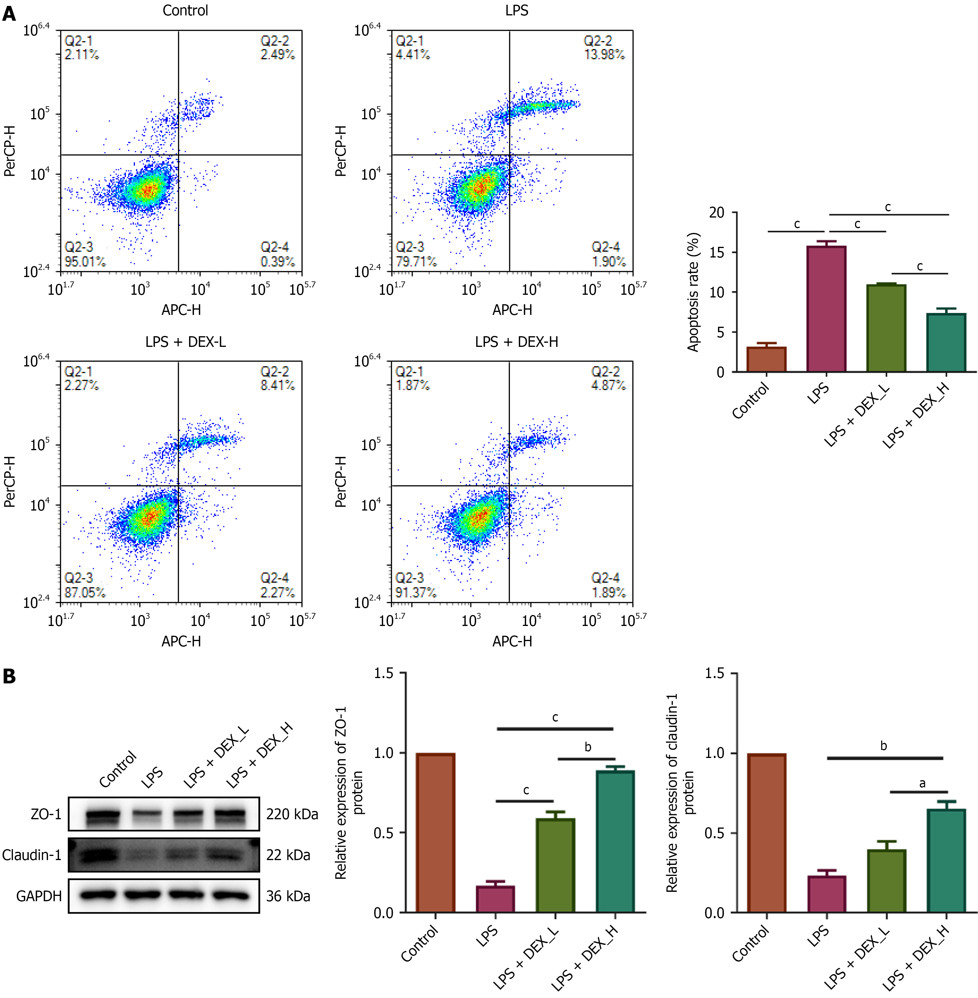
Figure 6 Dexmedetomidine attenuated lipopolysaccharide-induced apoptosis and upregulated the expression of tight junction proteins in IEC-6 cells.
A: Attenuated lipopolysaccharide-induced apoptosis; B: Upregulated the expression of tight junction proteins. aP < 0.01. bP < 0.001. cP < 0.0001. Dexmedetomidine (DEX)-low = 2.4 μM DEX; DEX-high = 4.8 μM DEX. Control group: IEC-6 cells maintained in standard culture medium; LPS group: IEC-6 cells treated with 5 μg/mL lipopolysaccharide for 72 hours; LPS + DEX-L group: IEC-6 cells pretreated with 2.4 μM dexmedetomidine for 24 hours followed by treatment with 5 μg/mL lipopolysaccharide for 72 hours; LPS + DEX-H group: IEC-6 cells pretreated with 4.8 μM dexmedetomidine for 24 hours followed by 5 μg/mL lipopolysaccharide for 72 hours; LPS: Lipopolysaccharide; DEX: Dexmedetomidine; L: Low; H: High; APC-H: Allophycocyanin-height; PerCP-H: Peridinin-chlorophyll-protein complex-height; GAPDH: Glyceraldehyde-3-phosphate dehydrogenase; ZO-1: Zonula occludens-1.
- Citation: Chen Y, Li CT, Wang JB, Tang WL, Zhao Y, Chen Y, Liao LM, Zhang LC, Lin TH, Cao ZF. Dexmedetomidine enhances anastomotic healing partly via the Wnt/β-catenin pathway in a rat model of colon surgery. World J Gastroenterol 2025; 31(44): 112833
- URL: https://www.wjgnet.com/1007-9327/full/v31/i44/112833.htm
- DOI: https://dx.doi.org/10.3748/wjg.v31.i44.112833