
















©The Author(s) 2025.
World J Gastroenterol. Nov 28, 2025; 31(44): 112187
Published online Nov 28, 2025. doi: 10.3748/wjg.v31.i44.112187
Published online Nov 28, 2025. doi: 10.3748/wjg.v31.i44.112187
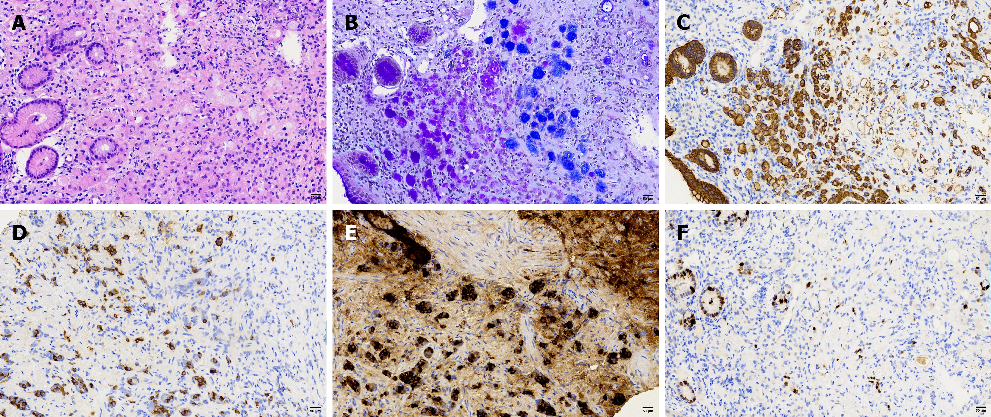
Figure 4 Pathological tissue strip #7.
The magnifications of all photographs are × 200. A: Signet-ring cell carcinoma cells, scale bar = 100 μm; B: Periodic acid Schiff-alcian blue (+), scale bar = 100 μm; C: Cytokeratin pan, scale bar = 100 μm; D: Signet-ring cell carcinoma cells were positive for synaptophysin, scale bar = 100 μm; E: Chromogranin A (+), scale bar = 100 μm; F: Ki-67 low expression, scale bar = 100 μm.
- Citation: Gao CZ, Meng Y, Du JZ, Zhu X. Very-early-stage gastric amphicrine carcinoma with mixed histology: A case report and review of literature. World J Gastroenterol 2025; 31(44): 112187
- URL: https://www.wjgnet.com/1007-9327/full/v31/i44/112187.htm
- DOI: https://dx.doi.org/10.3748/wjg.v31.i44.112187