
















©The Author(s) 2025.
World J Gastroenterol. Nov 21, 2025; 31(43): 111906
Published online Nov 21, 2025. doi: 10.3748/wjg.v31.i43.111906
Published online Nov 21, 2025. doi: 10.3748/wjg.v31.i43.111906
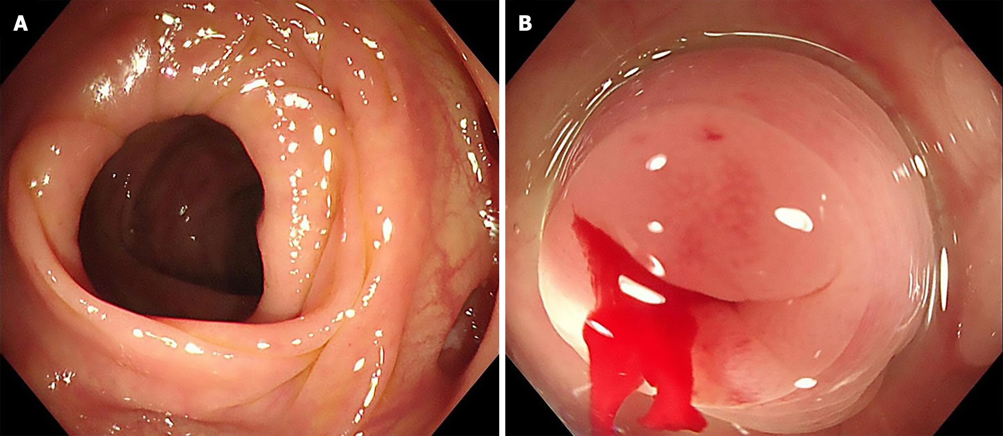
Figure 1 Endoscopic images of a colonic diverticulum.
A: Colonoscopy identified a diverticulum in the cecum; B: Repeat colonoscopy confirmed active bleeding originating from a diverticulum.
- Citation: Han TJ, Li JS. Utilizing the EyeMax direct visualization system for the treatment of colonic diverticular bleeding: A case report. World J Gastroenterol 2025; 31(43): 111906
- URL: https://www.wjgnet.com/1007-9327/full/v31/i43/111906.htm
- DOI: https://dx.doi.org/10.3748/wjg.v31.i43.111906