
















©The Author(s) 2025.
World J Gastroenterol. Nov 14, 2025; 31(42): 112354
Published online Nov 14, 2025. doi: 10.3748/wjg.v31.i42.112354
Published online Nov 14, 2025. doi: 10.3748/wjg.v31.i42.112354
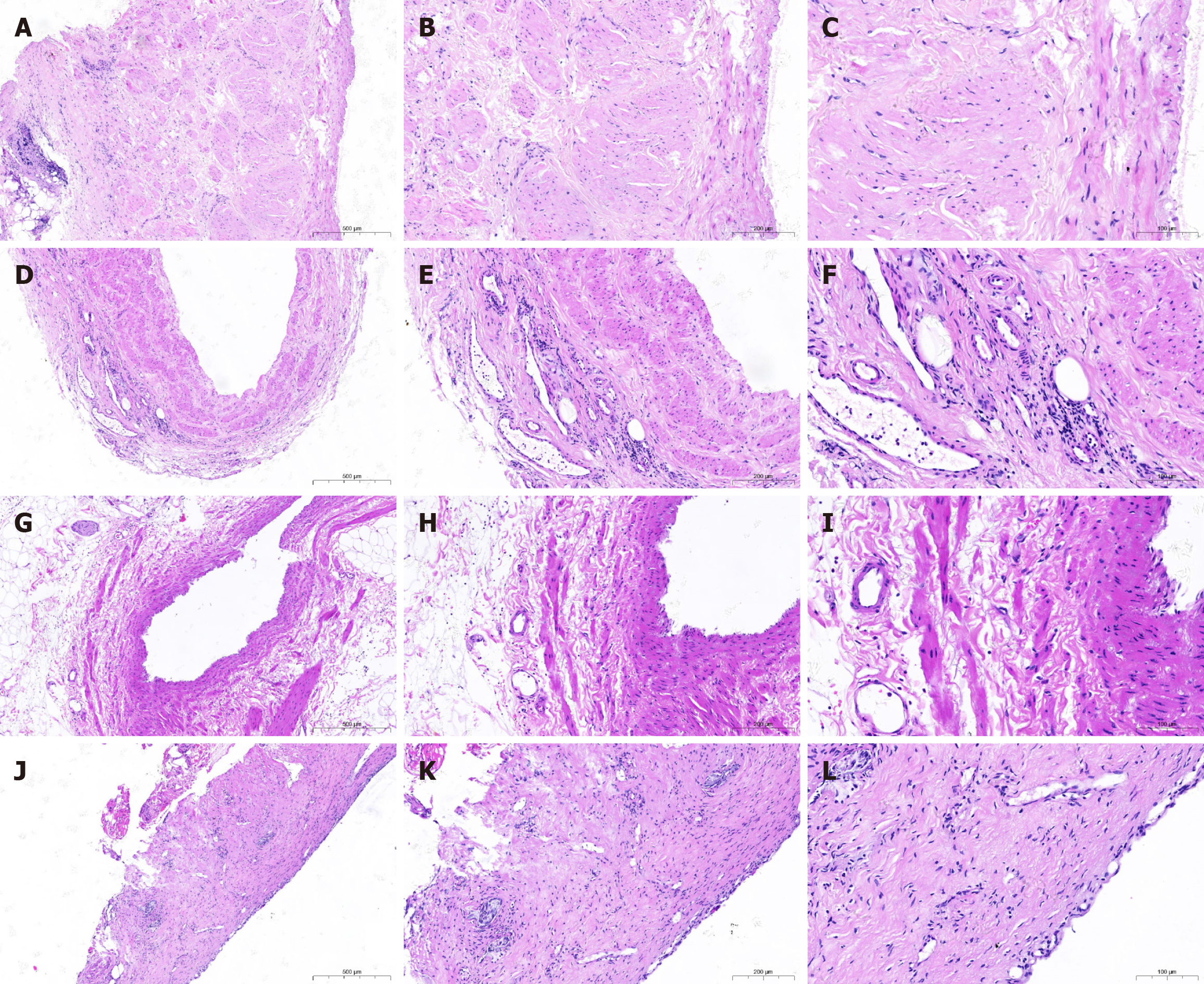
Figure 1 Hematoxylin and eosin staining.
A-C: Microscopic appearance of no portal-systemic venous invasion; D-F: Microscopic appearance of portal-systemic venous invasion into the adventitia; G-I: Microscopic appearance of portal-systemic venous invasion into the muscularis propria; J-L: Microscopic appearance of portal-systemic venous invasion into the intimal. A, D, G, and J: 100 ×; B, E, H, and K: 200 ×; C, F, I, and L: 400 ×.
- Citation: Wang FF, Dai XD, Zhao X, He Q, Lyu SC. Development and validation of a predictive model for portal-systemic venous invasion grading in borderline resectable pancreatic cancer. World J Gastroenterol 2025; 31(42): 112354
- URL: https://www.wjgnet.com/1007-9327/full/v31/i42/112354.htm
- DOI: https://dx.doi.org/10.3748/wjg.v31.i42.112354