















©The Author(s) 2025.
World J Gastroenterol. Nov 14, 2025; 31(42): 112196
Published online Nov 14, 2025. doi: 10.3748/wjg.v31.i42.112196
Published online Nov 14, 2025. doi: 10.3748/wjg.v31.i42.112196
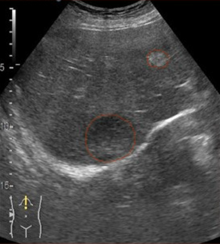
Figure 2 Representative contrast-enhanced ultrasound B-mode frame with expert lesion annotation.
The grayscale B-mode ultrasound image shows a focal liver lesion (outlined by the expert’s manual annotation in the B-mode field) that serves as the segmentation target. The corresponding binary mask (segmentation label) highlights the lesion region (distinguished from normal tissue) and provides the ground truth for training the deep learning model.
- Citation: Ciocalteu A, Urhut CM, Streba CT, Kamal A, Mamuleanu M, Sandulescu LD. Artificial intelligence in contrast enhanced ultrasound: A new era for liver lesion assessment. World J Gastroenterol 2025; 31(42): 112196
- URL: https://www.wjgnet.com/1007-9327/full/v31/i42/112196.htm
- DOI: https://dx.doi.org/10.3748/wjg.v31.i42.112196