















©The Author(s) 2025.
World J Gastroenterol. Nov 14, 2025; 31(42): 112196
Published online Nov 14, 2025. doi: 10.3748/wjg.v31.i42.112196
Published online Nov 14, 2025. doi: 10.3748/wjg.v31.i42.112196
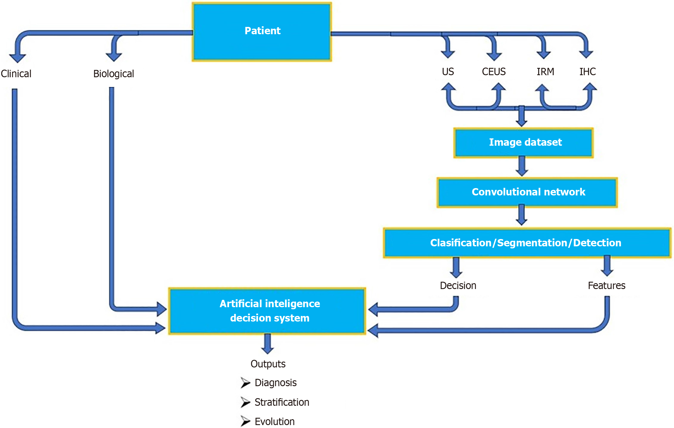
Figure 1 Artificial intelligence-driven decision workflow integrating contrast-enhanced ultrasound and multimodal data.
This diagram illustrates an integrated clinical workflow in which artificial intelligence (AI) supports decision-making in hepatocellular carcinoma assessment. The patient contributes multimodal data (clinical, biological and imaging) acquired through conventional ultrasound, contrast-enhanced ultrasound (CEUS), magnetic resonance imaging and pathology results from immunohistochemistry (an antibody-based tissue staining method that highlights specific disease markers). These imaging datasets are processed through a convolutional neural network model as previously described in the “Machine learning and deep learning architectures for CEUS analysis” section, enabling lesion classification, segmentation, and detection within the integrated decision workflow. The resulting features and decisions are input into an AI decision system, which synthesizes information and generates clinically actionable outputs, including diagnosis, risk stratification, and disease evolution prediction. US: Ultrasound; CEUS: Contrast-enhanced ultrasound.
- Citation: Ciocalteu A, Urhut CM, Streba CT, Kamal A, Mamuleanu M, Sandulescu LD. Artificial intelligence in contrast enhanced ultrasound: A new era for liver lesion assessment. World J Gastroenterol 2025; 31(42): 112196
- URL: https://www.wjgnet.com/1007-9327/full/v31/i42/112196.htm
- DOI: https://dx.doi.org/10.3748/wjg.v31.i42.112196