
















©The Author(s) 2025.
World J Gastroenterol. Nov 14, 2025; 31(42): 111706
Published online Nov 14, 2025. doi: 10.3748/wjg.v31.i42.111706
Published online Nov 14, 2025. doi: 10.3748/wjg.v31.i42.111706
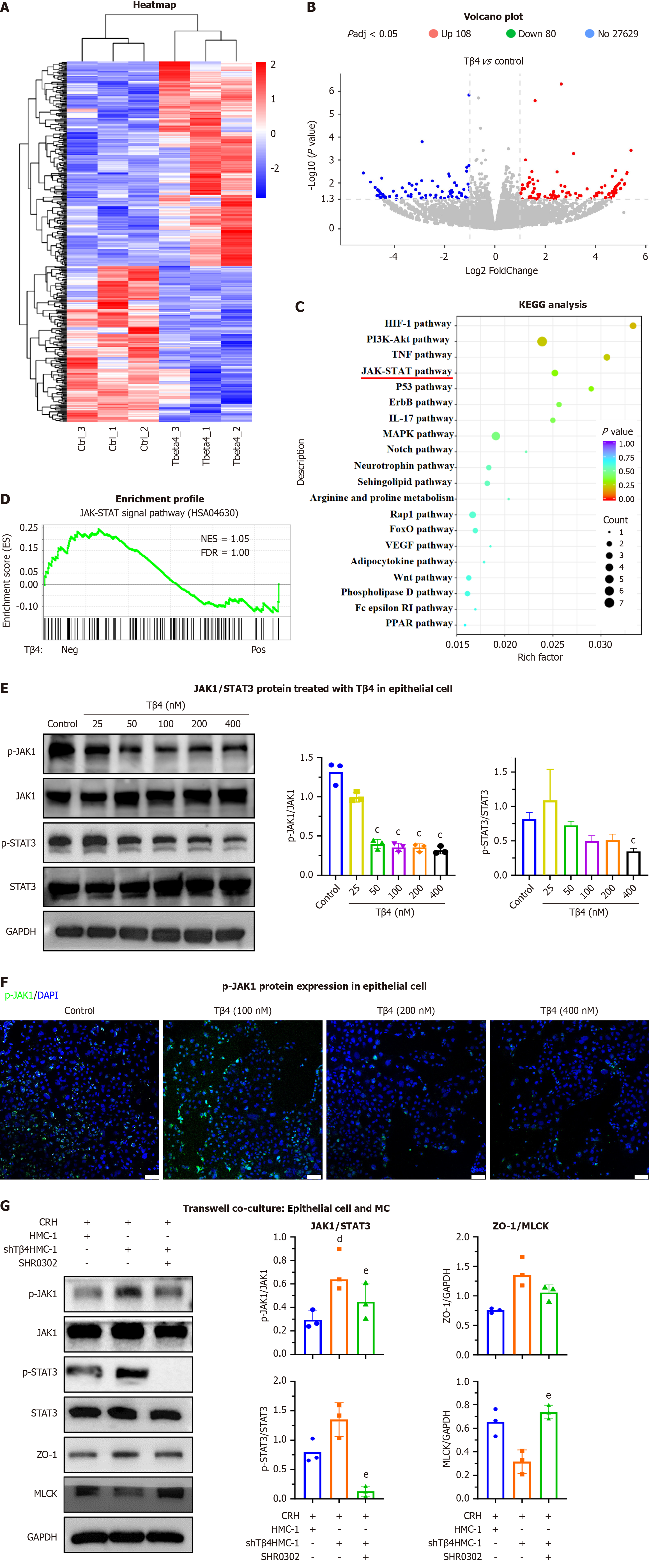
Figure 9 Thymosin β4 regulates the Janus kinase 1/signal transducer and activation of transcription 3 pathways in epithelial cells.
Caco2 cells were treated with thymosin β4 (Tβ4). A: Heatmap of differentially expressed mRNAs; B: Volcano plot of RNA-seq results (n = 3 biological replicates/group; differential expression defined as |log2 (fold change)| > 1 and FDR < 0.05); C: Top 20 enriched pathways by KEGG analysis; D: GSEA analysis of Janus kinase 1 (JAK1)/signal transducer and activation of transcription 3 (STAT3) pathway enrichment in Tβ4-positive vs -negative Caco2 cell; E: Representative western blot images and biochemical quantification of p-JAK1, JAK1, p-STAT3, STAT3. Bar, SD, cP < 0.05 vs Control; F: Immunofluorescence staining of p-JAK1 in Caco2 cells. Scale bar, 100 μm; G: Activated mast cells [HMC-1 and HMC-1shTβ4 treated with corticotropin-releasing hormone (CRH)] stimulated with SHR0302 and co-cultured with Caco2 cells, and representative western blot images and biochemical quantification of p-JAK1, JAK1, p-STAT3, STAT3, zonulin 1, myosin light chain kinase and GAPDH. Bar, SD, dP < 0.05 vs “HMC-1 with CRH”, eP < 0.05 vs “HMC-1shTβ4 with CRH”. Tβ4: Thymosin β4; JAK1: Janus kinase 1; STAT3: Signal transducer and activation of transcription 3; MC: Mast cell; ZO-1: Zonulin 1; MLCK: Myosin light chain kinase; CRH: Corticotropin-releasing hormone.
- Citation: Sun YS, Bai XQ, Sun KD, Li J, Liu L, Chen YY, Zeng ZY, Wang Q, Guo YB. Thymosin β4 released by mast cells under stress conditions impairs intestinal epithelial barrier via IL22RA1/JAK1/STAT3 signaling in irritable bowel syndrome. World J Gastroenterol 2025; 31(42): 111706
- URL: https://www.wjgnet.com/1007-9327/full/v31/i42/111706.htm
- DOI: https://dx.doi.org/10.3748/wjg.v31.i42.111706