
















©The Author(s) 2025.
World J Gastroenterol. Nov 14, 2025; 31(42): 111706
Published online Nov 14, 2025. doi: 10.3748/wjg.v31.i42.111706
Published online Nov 14, 2025. doi: 10.3748/wjg.v31.i42.111706
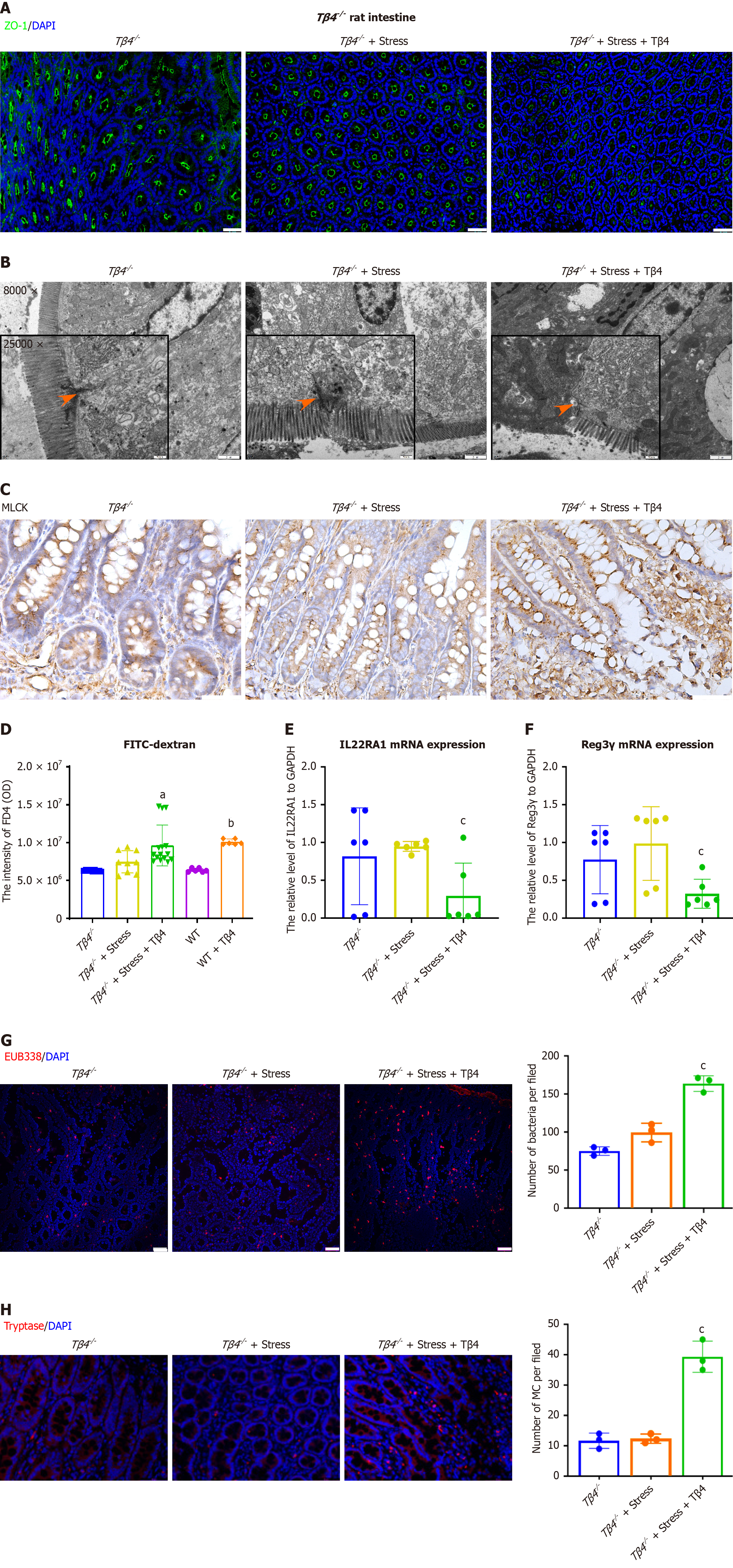
Figure 5 Thymosin β4 deficiency protects rats from stress-induced injury in the intestine.
Thymosin β4 (Tβ4) (3 mg/kg) was injected into stress-induced Tβ4 deficiency (Tβ4−/−) rats. A: Representative intestinal immunofluorescence images of zonulin 1. Scale bar, 50 μm; B: Representative TEM images of the colon. Magnification: 8000 ×, Scale bar, 2 μm. Magnification: 25000 ×, Scale bar, 500 nm; C: Immunohistochemistry staining of myosin light chain kinase. Scale bar, 100 μm; D: FITC-dextran 4000 (FD4, 4 kDa, 5 mg/kg) was orally administered, and the flux of FITC-dextran in rat serum was observed 3 hours later. aP < 0.05 vs the Tβ4−/− group, bP < 0.001 vs the WT group; E and F: The colonic tissue mRNA levels of (E) IL22RA1 and (F) Reg3γ. Bar, SD, cP < 0.05 vs the Tβ4−/− under stress group; G: Representative FISH staining for bacterial infiltration using EUB338 probe (red) and DAPI (blue). Scale bar, 50 μm. Bar, SD, cP < 0.05 vs the Tβ4−/− under stress group; H: Representative intestinal immunofluorescence images of mast cell (Tryptase, red) and DAPI (blue), Scale bar, 50 μm. Bar, SD, cP < 0.05 vs the Tβ4−/− under stress group. MLCK: Myosin light chain kinase; Tβ4: Thymosin β4; IL22RA1: Interleukin 22 receptor A1; WT: Wild-type.
- Citation: Sun YS, Bai XQ, Sun KD, Li J, Liu L, Chen YY, Zeng ZY, Wang Q, Guo YB. Thymosin β4 released by mast cells under stress conditions impairs intestinal epithelial barrier via IL22RA1/JAK1/STAT3 signaling in irritable bowel syndrome. World J Gastroenterol 2025; 31(42): 111706
- URL: https://www.wjgnet.com/1007-9327/full/v31/i42/111706.htm
- DOI: https://dx.doi.org/10.3748/wjg.v31.i42.111706