
















©The Author(s) 2025.
World J Gastroenterol. Nov 14, 2025; 31(42): 111706
Published online Nov 14, 2025. doi: 10.3748/wjg.v31.i42.111706
Published online Nov 14, 2025. doi: 10.3748/wjg.v31.i42.111706
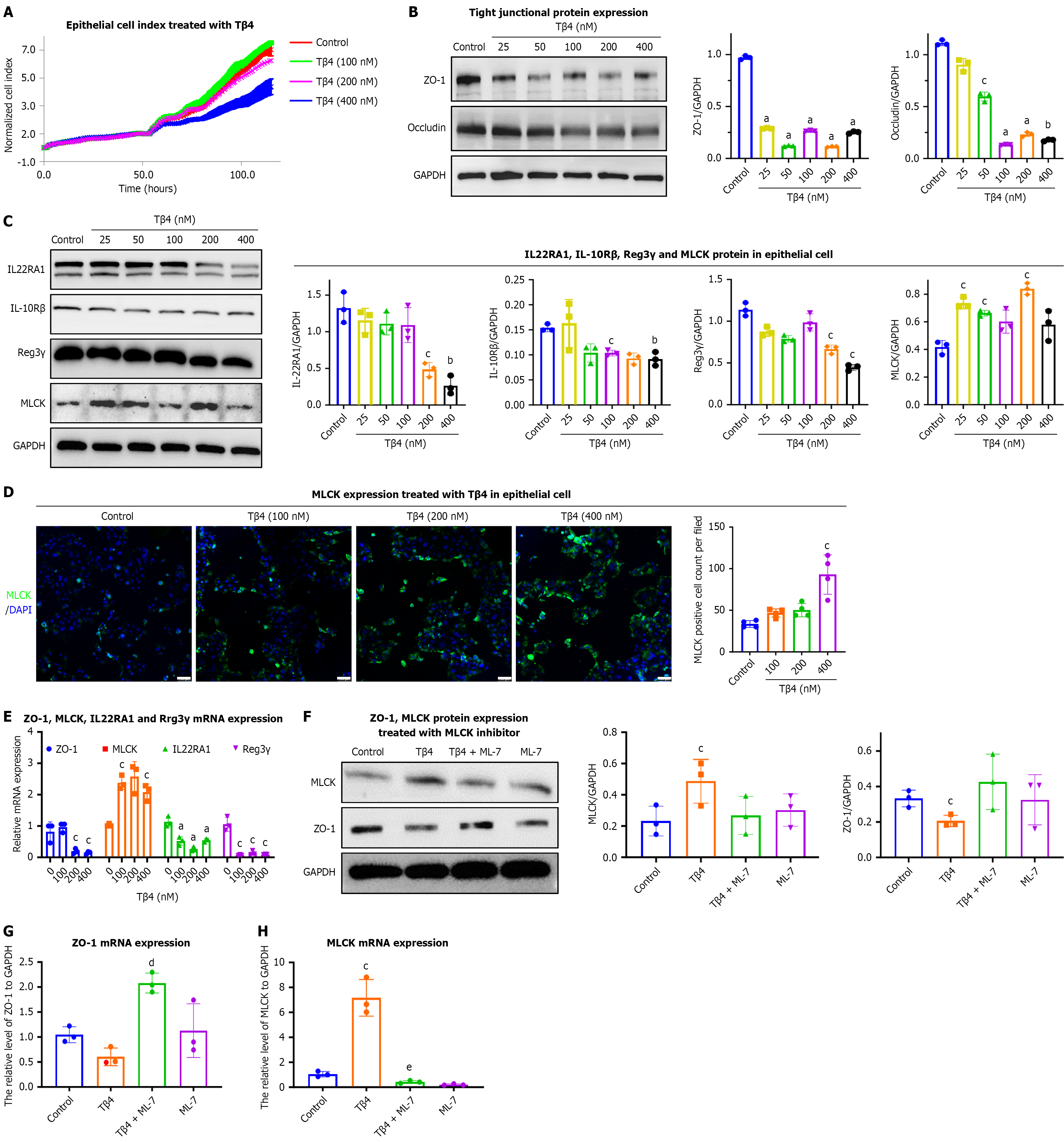
Figure 3 Thymosin β4 is sufficient to drive intestinal permeability and dysbiosis in vitro.
A: The epithelial barrier function of Caco2 was measured after treatment with thymosin β4 (Tβ4) (100 nmol/L, 200 nmol/L, 400 nmol/L). Each line represents the mean of 3 wells per condition. The cell index serves as a real-time value reflecting changes in barrier properties; B and C: Caco2 cells were treated with Tβ4 (25 nmol/L, 50 nmol/L, 100 nmol/L, 200 nmol/L, 400 nmol/L) for 24 hours, and the protein levels of (B) zonulin 1 (ZO-1), Occludin, (C) interleukin 22 receptor A1 (IL22RA1), IL-10Rβ, Reg3γ, and myosin light chain kinase (MLCK) were detected by western blot assay. Bar, SD, aP < 0.001, bP < 0.01, cP < 0.05 vs the Control group; D: Representative immunofluorescence images of MLCK (green) and DAPI (blue). Scale bar, 100 μm. Bar, SD, cP < 0.05 vs the Control group; E: After treatment with Tβ4 (100 nmol/L, 200 nmol/L, 400 nmol/L) for 24 hours, the mRNA levels of ZO-1, IL22RA1, MLCK, and Reg3γ were evaluated by qPCR. Bar, SD, aP < 0.001, cP < 0.05 vs the Control group (0 nmol/L); F: Cells were treated with Tβ4 (200 nmol/L) for 24 h in MLCK inhibitor (ML-7)-induced Caco2 (prepared 30 minutes in advance), and then the protein levels of MLCK and ZO-1 were evaluated by western blotting; G and H: After treatment with ML-7 in Tβ4-treated Caco2, the mRNA level of (G) ZO-1 and (H) MLCK were evaluated by qPCR. Bar, SD, cP < 0.05 vs the Control group, dP < 0.001 vs the Tβ4 group, eP < 0.05 vs the Tβ4 group. MLCK: Myosin light chain kinase; ZO-1: Zonulin 1; Tβ4: Thymosin β4; IL: Interleukin; IL-10R: Interleukin-10 receptor.
- Citation: Sun YS, Bai XQ, Sun KD, Li J, Liu L, Chen YY, Zeng ZY, Wang Q, Guo YB. Thymosin β4 released by mast cells under stress conditions impairs intestinal epithelial barrier via IL22RA1/JAK1/STAT3 signaling in irritable bowel syndrome. World J Gastroenterol 2025; 31(42): 111706
- URL: https://www.wjgnet.com/1007-9327/full/v31/i42/111706.htm
- DOI: https://dx.doi.org/10.3748/wjg.v31.i42.111706