
















©The Author(s) 2025.
World J Gastroenterol. Nov 7, 2025; 31(41): 112794
Published online Nov 7, 2025. doi: 10.3748/wjg.v31.i41.112794
Published online Nov 7, 2025. doi: 10.3748/wjg.v31.i41.112794
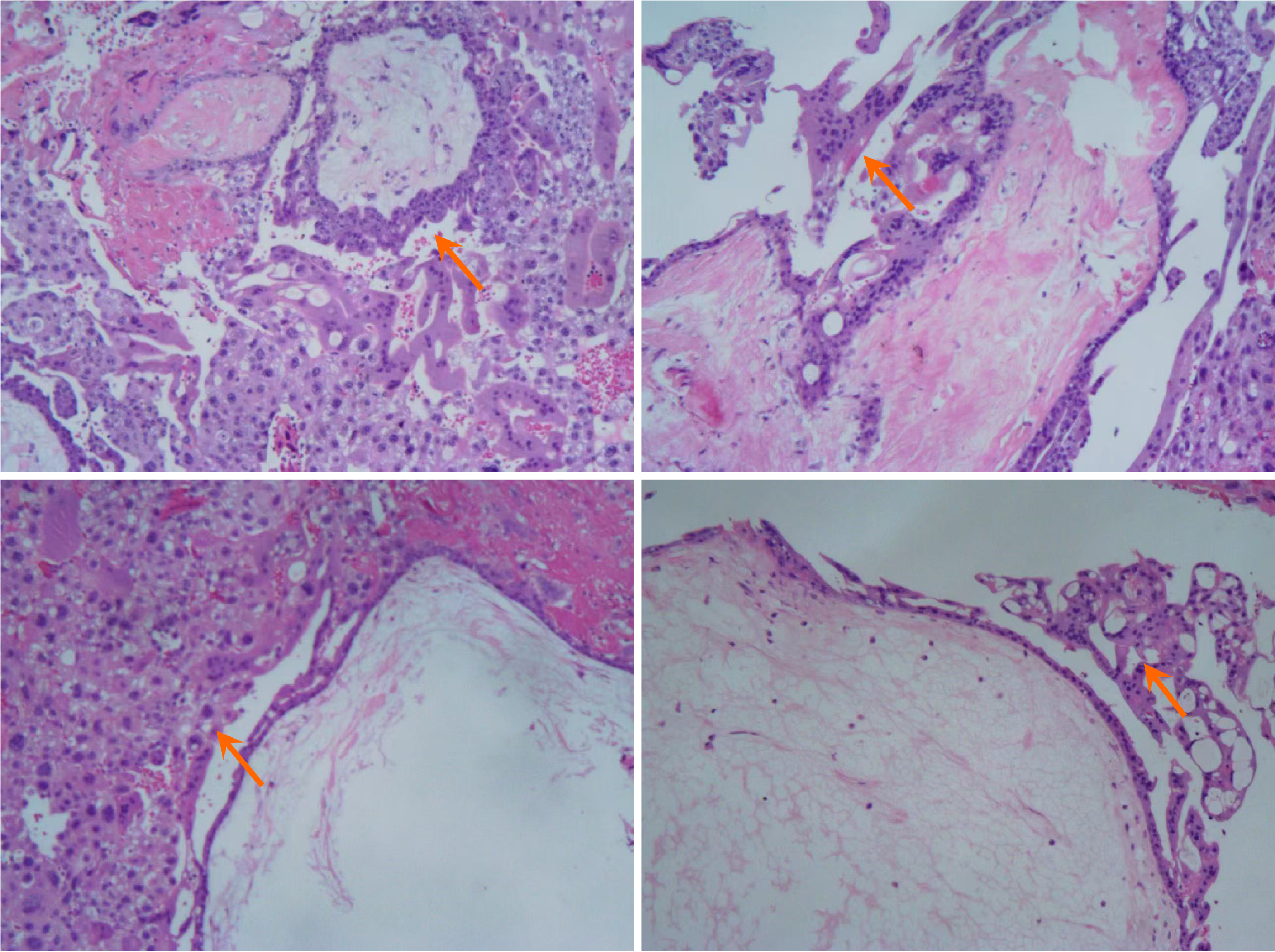
Figure 6 On the histopathology of the resected specimen, the hemorrhage, necrosis, and hyperplasia of trophoblastic cells were ob
- Citation: Yu WH, Zong Y, Zhou JW, Xu GQ. Uncommon causes of acute small intestinal bleeding-invasive mole: A case report and review of literature. World J Gastroenterol 2025; 31(41): 112794
- URL: https://www.wjgnet.com/1007-9327/full/v31/i41/112794.htm
- DOI: https://dx.doi.org/10.3748/wjg.v31.i41.112794