
















©The Author(s) 2025.
World J Gastroenterol. Nov 7, 2025; 31(41): 111449
Published online Nov 7, 2025. doi: 10.3748/wjg.v31.i41.111449
Published online Nov 7, 2025. doi: 10.3748/wjg.v31.i41.111449
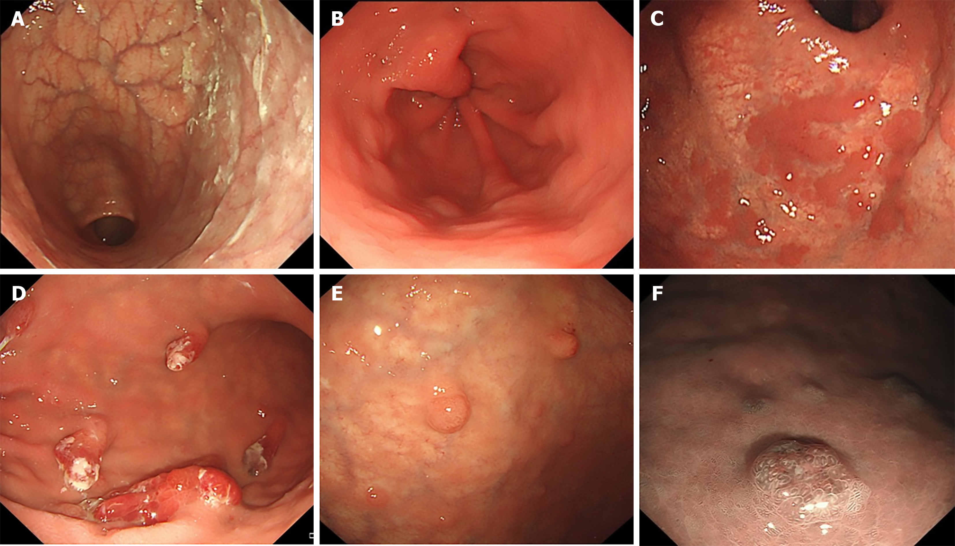
Figure 1 Some endoscopic findings in patients with autoimmune gastritis.
A: Mucosal atrophy of the gastric fundus and corpus (reverse atrophy) with sticky adherent dense mucus; B: Circular wrinkle-like pattern of the mucosa with no atrophy in antrum; C: Remnants of oxyntic mucosa, presenting as flat localized type; D: Hyperplastic polyps; E: Scattered type I gastric neuroendocrine tumors in the atrophic gastric fundus and body; F: Narrow band imaging of gastric neuroendocrine tumors.
- Citation: Li YM, Guo WJ, Deng C, Luo J, Shi YF, Zhu D, Wei QL, Zhang MG, Du SY, Tan HY. Risk assessment of type I gastric neuroendocrine tumors based on endoscopic and clinical features of autoimmune gastritis. World J Gastroenterol 2025; 31(41): 111449
- URL: https://www.wjgnet.com/1007-9327/full/v31/i41/111449.htm
- DOI: https://dx.doi.org/10.3748/wjg.v31.i41.111449