
















©The Author(s) 2025.
World J Gastroenterol. Nov 7, 2025; 31(41): 111174
Published online Nov 7, 2025. doi: 10.3748/wjg.v31.i41.111174
Published online Nov 7, 2025. doi: 10.3748/wjg.v31.i41.111174
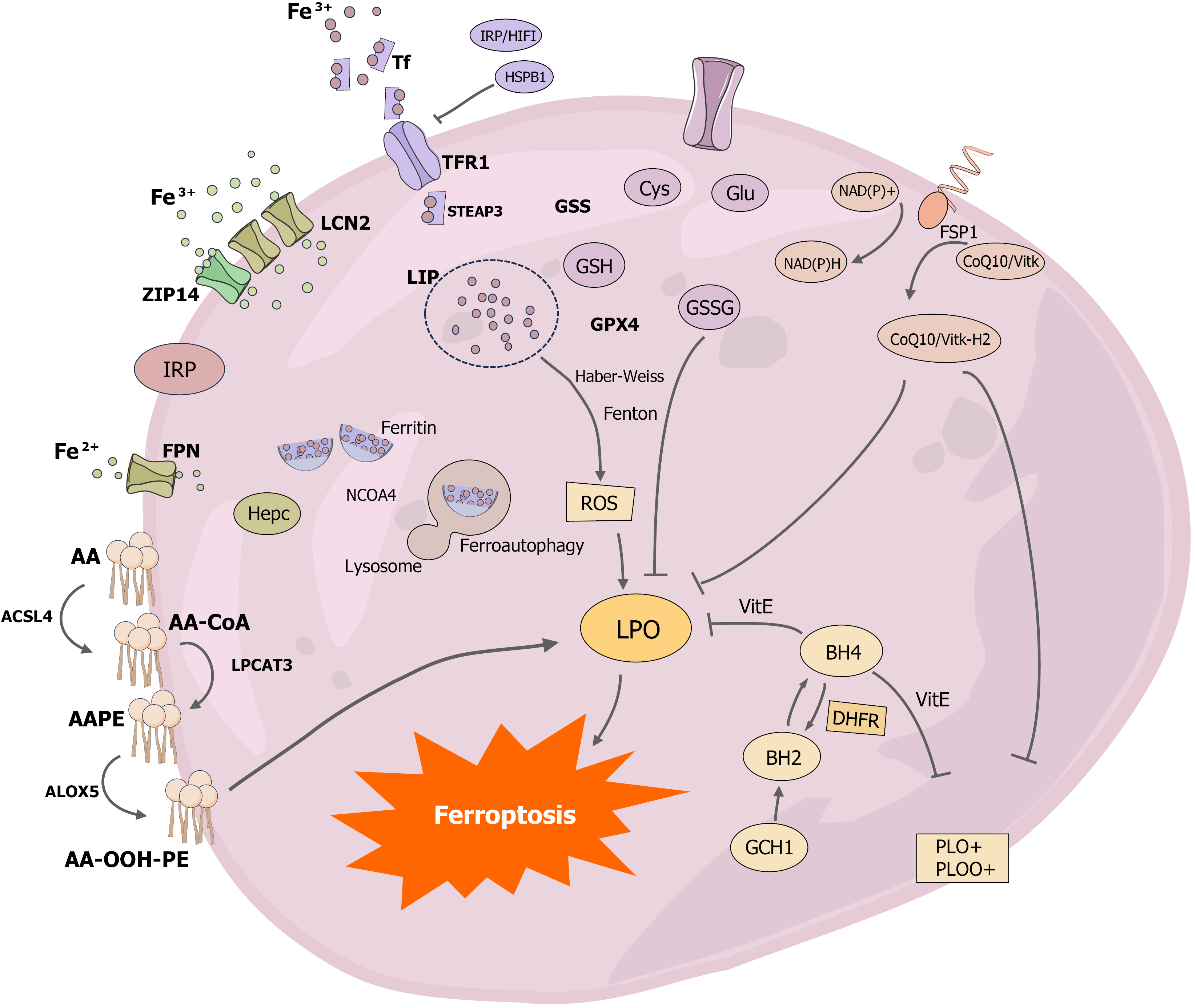
Figure 2 Diagram of the mechanism of development of ferroptosis.
The key molecular mechanisms of ferroptosis have been systematically investigated, with a focus on understanding the pathways and mechanisms of iron-related processes. Ferritin, the primary storage protein for iron in the body, plays a central role in iron metabolism. Iron is primarily acquired in the bloodstream as Fe3+, which enters cells through the transferrin receptor 1 (TFR1) on the cell surface. The expression of TFR1 is regulated by iron regulatory protein and hypoxia-inducible factor-1, while heat shock protein B1 inhibits TFR1 expression. Fe3+ is reduced to Fe2+ by the lipid carrier protein 2, which allows free iron to enter the cytoplasmic iron-unstable pools. Additionally, ZRT/IRT-like proteins such as ZRT/IRT-like protein 14 can also mediate iron entry into the cell. Ferritin, the sole channel protein for Fe2+ export, is primarily regulated by hemosiderin, and ferritin autophagy is a critical mechanism for maintaining intracellular free Fe2+ levels. When autophagy receptor nuclear coactivator 4 is present, ferritin binds to autophagosomes, increasing intracellular free Fe2+ levels. Free iron in the cytoplasmic iron-unstable pools generates reactive oxygen species through the Fenton reaction and Haber-Weiss reaction. Enzymatic pathways, such as long-chain family 4 catalyzing arachidonoyl-CoA formation, lysophosphatidylcholine acyltransferase 3 controlling the esterification of arachidonoyl-CoA to arachidonoyl-phosphatidylethanolamine, and arachidonic acid lipoxygenases oxidizing arachidonoyl-phosphatidylethanolamine to AA-OOH-PE, contribute to the generation of reactive oxygen species. The primary antioxidant pathways in ferroptosis include the cystine/glutamate antiporter/glutathione peroxidase 4/glutathione axis, the ferroptosis suppressor protein 1-CoQ10-NAD(P)H axis, and the GTP cyclohydrolase 1-dihydrofolate reductase-BH4 axis. Notably, the ferroptosis suppressor protein 1-CoQ10-NAD(P)H axis and the GTP cyclohydrolase 1-dihydrofolate reductase-BH4 axis function to inhibit fatty acid synthesis, thereby contributing to the oxidative stress response in ferroptosis. Tf: Transferrin; IRP: Iron regulatory protein; HIF1: Hypoxia-inducible factor 1; HSPB1: Heat shock protein B1; TFR1: Transferrin receptor 1; LCN2: Lipid carrier protein 2; GSS: Glutathione synthetase; ZIP14: ZRT/IRT-like protein 14; GSH: Glutathione; GSSG: Glutathione disulfide; FSP1: Ferroptosis suppressor protein 1; GPX4: Glutathione peroxidase 4; FPN: Ferroportin; ROS: Reactive oxygen species; AA: Arachidonic acid; AA-CoA: Arachidonoyl-CoA; AA-PE: Arachidonoyl-phosphatidylethanolamine; ACSL4: Acyl coenzyme A synthetase long-chain family 4; LPCAT3: Lyso-phosphatidylcholine acyltransferase-3; ALOX5: Arachidonic acid lipoxygenases 5; LPO: Lipid peroxidation; DHFR: Dihydrofolate reductase; GCH1: GTP cyclohydrolase 1.
- Citation: Han JF, Jia ZY, Fan X, Zhao XY, Cheng LY, Xia YX, Ji XR, Zang WQ. Mechanisms of ferroptosis in primary hepatocellular carcinoma and progress of artificial intelligence-based predictive modeling in hepatocellular carcinoma. World J Gastroenterol 2025; 31(41): 111174
- URL: https://www.wjgnet.com/1007-9327/full/v31/i41/111174.htm
- DOI: https://dx.doi.org/10.3748/wjg.v31.i41.111174