
















©The Author(s) 2025.
World J Gastroenterol. Nov 7, 2025; 31(41): 110398
Published online Nov 7, 2025. doi: 10.3748/wjg.v31.i41.110398
Published online Nov 7, 2025. doi: 10.3748/wjg.v31.i41.110398
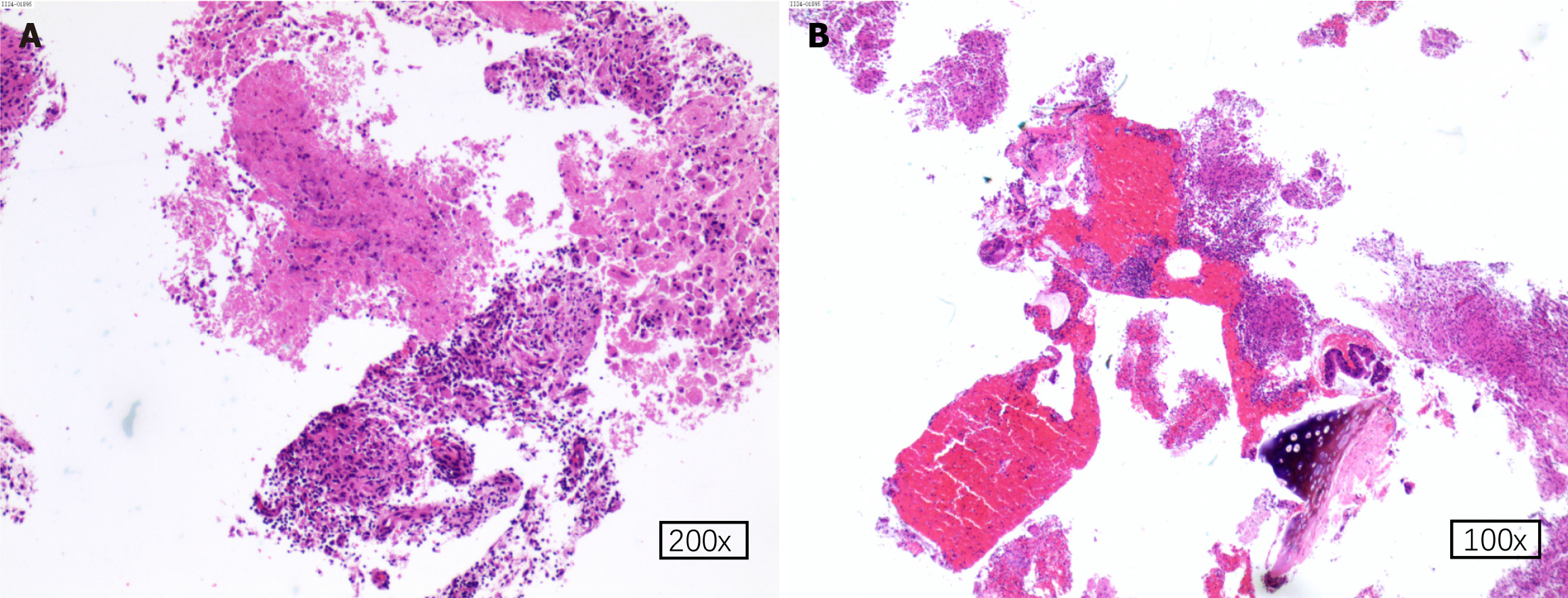
Figure 4 Histopathological examination.
A: Granulomatous inflammation with central necrosis is observed in focal areas. Occasional mucinous epithelial cells with preserved differentiation are also identified (hematoxylin and eosin staining, magnification × 200); B: Microscopic examination showing scattered lymphocytes, histiocytes, and cartilage fragments (hematoxylin and eosin staining, magnification × 100).
- Citation: Nima CL, Wang HG, Zhou Q. Pancreatic tuberculosis: A case report and review of literature. World J Gastroenterol 2025; 31(41): 110398
- URL: https://www.wjgnet.com/1007-9327/full/v31/i41/110398.htm
- DOI: https://dx.doi.org/10.3748/wjg.v31.i41.110398