
















©The Author(s) 2025.
World J Gastroenterol. Nov 7, 2025; 31(41): 110398
Published online Nov 7, 2025. doi: 10.3748/wjg.v31.i41.110398
Published online Nov 7, 2025. doi: 10.3748/wjg.v31.i41.110398
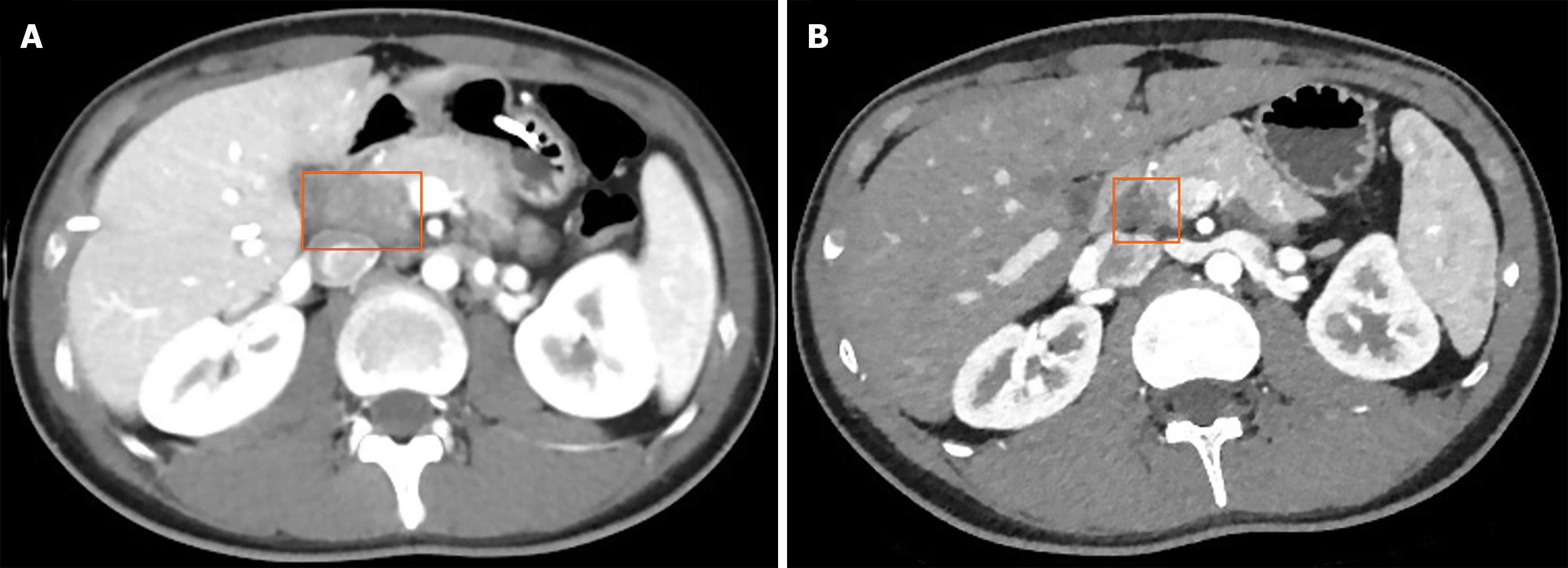
Figure 1 Abdominal contrast-enhanced computed tomography images.
A: An irregular hypodense mass located in the duodenum and pancreatic head, measuring approximately 29.8 mm × 22.0 mm (orange box), with multiple internal air-fluid levels. The mass demonstrates peripheral contrast enhancement and indistinct surrounding fat planes, indicating local inflammatory infiltration or possible extension; B: Follow-up imaging shows a mildly hypodense mass in the duodenal-pancreatic head region, reduced in size compared to the prior scan (December 25, 2023). Previously observed gas and fluid components have been absorbed (orange box).
- Citation: Nima CL, Wang HG, Zhou Q. Pancreatic tuberculosis: A case report and review of literature. World J Gastroenterol 2025; 31(41): 110398
- URL: https://www.wjgnet.com/1007-9327/full/v31/i41/110398.htm
- DOI: https://dx.doi.org/10.3748/wjg.v31.i41.110398