
















©The Author(s) 2025.
World J Gastroenterol. Oct 21, 2025; 31(39): 110115
Published online Oct 21, 2025. doi: 10.3748/wjg.v31.i39.110115
Published online Oct 21, 2025. doi: 10.3748/wjg.v31.i39.110115
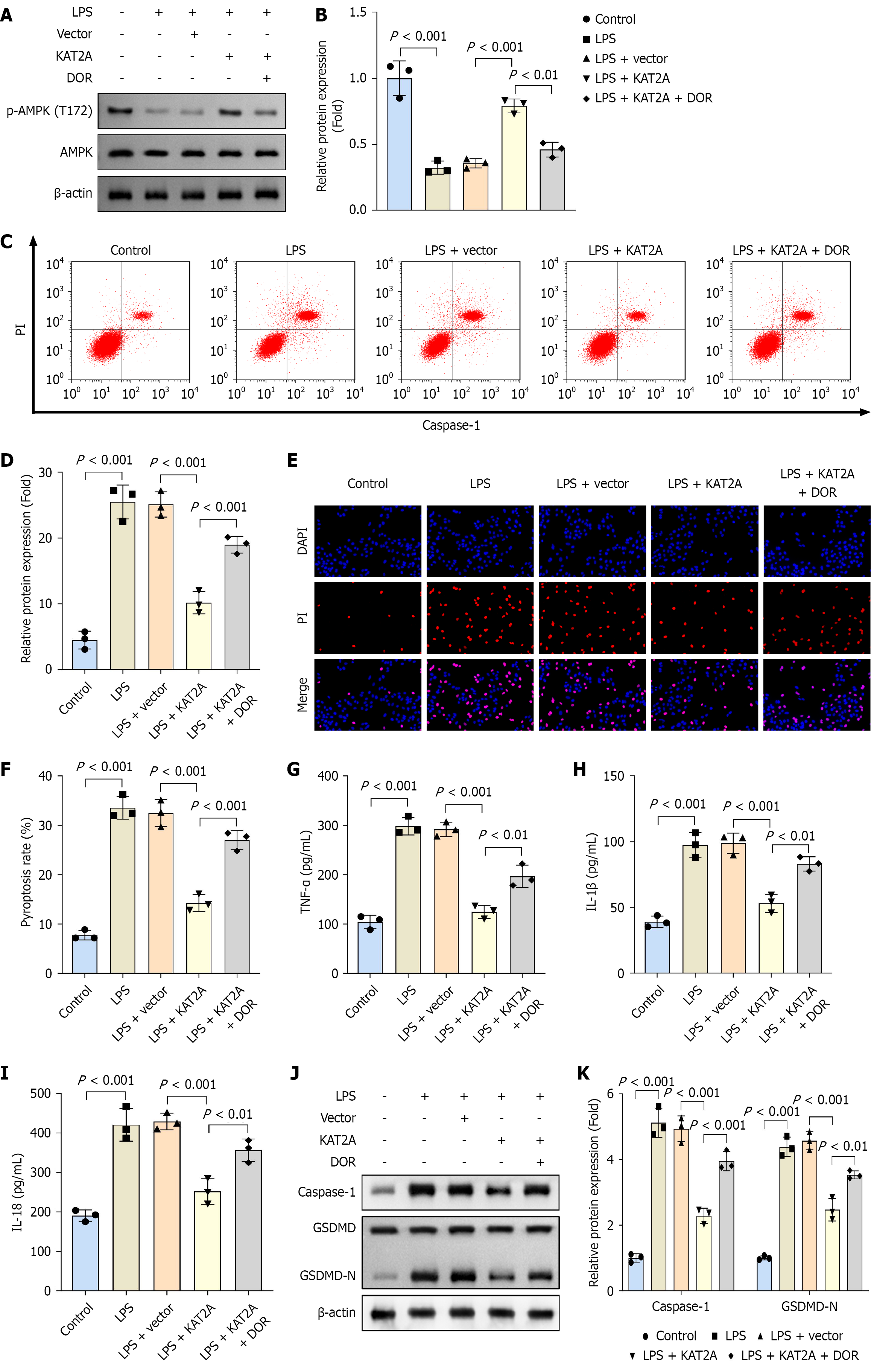
Figure 5 Dorsomorphin treatment increased pyroptosis and inflammation in gallbladder mucosal epithelial cells.
A: Western blot was performed to analyze the protein level of phospho-adenosine monophosphate-activated protein kinase (AMPK) (p-AMPK) (T172)/AMPK in gallbladder mucosal epithelial cells (GMECs) (n = 3); B: Quantification of relative protein level of p-AMPK (T172)/AMPK in each group (n = 3); C and D: Flow cytometry was performed to assess the pyroptosis rate in GMECs in each group (n = 3); E and F: Propidium iodide staining was used to analyze cell death in each group (n = 3); G: Enzyme-linked immunosorbent assay was used to measure the concentrations of tumor necrosis factor-α; H: Interleukin (IL)-1β; I: IL-18 in each group (n = 3); J and K: Western blot was performed to assess the protein levels of caspase-1 and gasdermin D-N in each group (n = 3). LPS: Lipopolysaccharide; DOR: Dorsomorphin.
- Citation: Wang XX, Ma MZ, Zhu LC, Dai LF, Qin C, Shao S, Xu XW, Gao RX, Zhang ZH. Lysine acetyltransferase 2A-mediated succinylation of adenosine monophosphate-activated protein kinase suppresses gallstone formation by inhibiting inflammation and pyroptosis. World J Gastroenterol 2025; 31(39): 110115
- URL: https://www.wjgnet.com/1007-9327/full/v31/i39/110115.htm
- DOI: https://dx.doi.org/10.3748/wjg.v31.i39.110115