
















©The Author(s) 2025.
World J Gastroenterol. Oct 21, 2025; 31(39): 110115
Published online Oct 21, 2025. doi: 10.3748/wjg.v31.i39.110115
Published online Oct 21, 2025. doi: 10.3748/wjg.v31.i39.110115
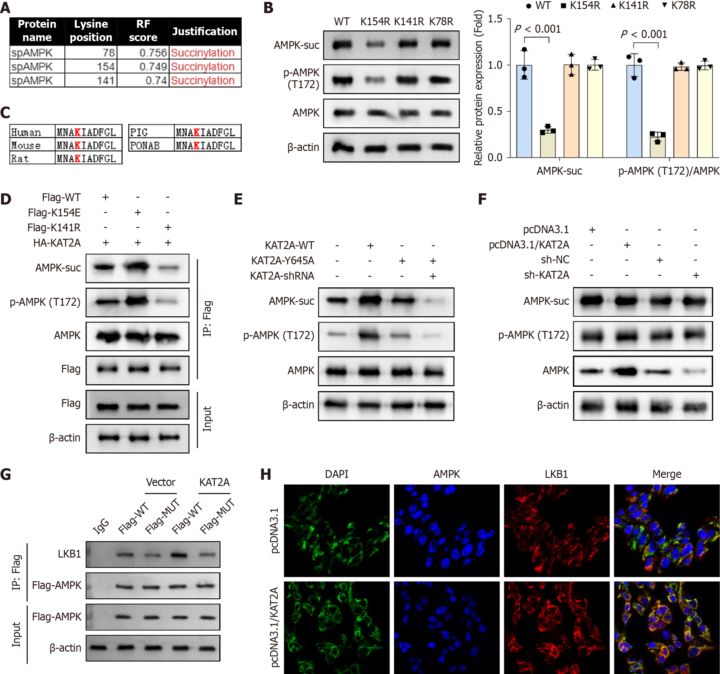
Figure 4 Adenosine monophosphate-activated protein kinase was succinylated at K170 site in gallbladder mucosal epithelial cells.
A: The protein levels of adenosine monophosphate-activated protein kinase (AMPK) succinylation (AMPK-suc), phospho-AMPK (p-AMPK), and AMPK after lysine acetyltransferase 2A (KAT2A) overexpression were detected by immunoprecipitation (IP) and western bot assays (n = 3); B: After mutating K179, K170, and K204 to K179R, K170R, and K204R, IP and western blot were performed to analyze the protein levels of AMPK-suc, p-AMPK, and AMPK (n = 3); C: K154 was highly conserved in different species, including human, mouse, rat, pig, and pongo abelii; D and E: Gallbladder mucosal epithelial cells (GMECs) were transfected with flag-WT, flag-K154E, flag-K154R, HA-KAT2A, KAT2A-WT, KAT2A-Y645A, and KAT2A-shRNA plasmids, and the protein levels of AMPK-suc and p-AMPK/AMPK was analyzed by IP and western blot (n = 3); F: After transfecting pcDNA3.1, pcDNA3.1/KAT2A, sh-KAT2A, and sh-NC plasmids, the protein levels of liver kinase B1 (LKB1)-suc, LKB1, and KAT2A were analyzed by IP and western blot (n = 3); G: Co-immunoprecipitation (Co-IP) was performed to detect the interaction between LKB1 and AMPK in GMECs (n = 3); H: Immunofluorescence (IF) was performed to analyze the binding of LKB1 and AMPK in GMECs (n = 3). DAPI: 4',6-diamidino-2-phenylindole.
- Citation: Wang XX, Ma MZ, Zhu LC, Dai LF, Qin C, Shao S, Xu XW, Gao RX, Zhang ZH. Lysine acetyltransferase 2A-mediated succinylation of adenosine monophosphate-activated protein kinase suppresses gallstone formation by inhibiting inflammation and pyroptosis. World J Gastroenterol 2025; 31(39): 110115
- URL: https://www.wjgnet.com/1007-9327/full/v31/i39/110115.htm
- DOI: https://dx.doi.org/10.3748/wjg.v31.i39.110115