
















©The Author(s) 2025.
World J Gastroenterol. Oct 14, 2025; 31(38): 111549
Published online Oct 14, 2025. doi: 10.3748/wjg.v31.i38.111549
Published online Oct 14, 2025. doi: 10.3748/wjg.v31.i38.111549
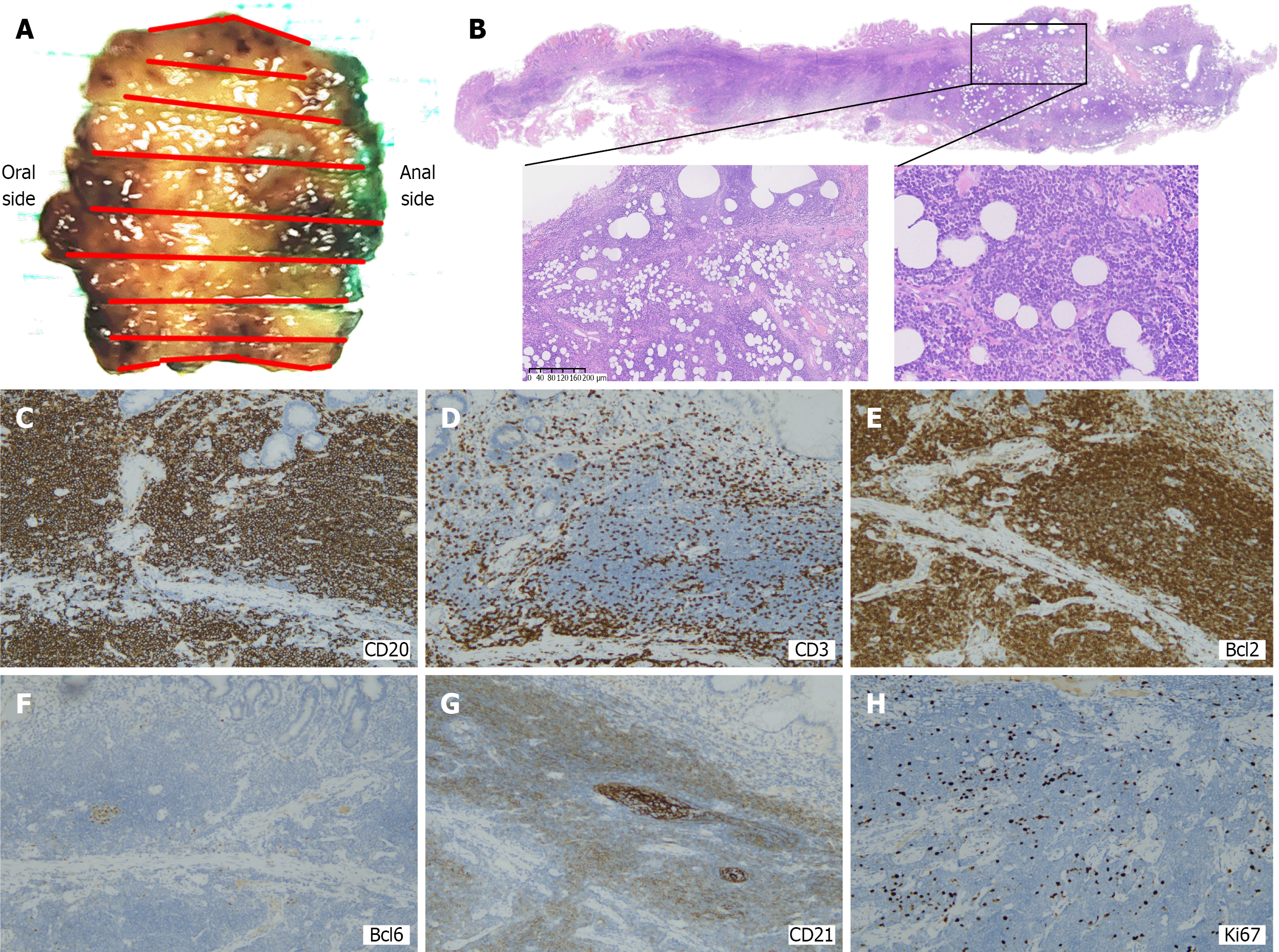
Figure 3 Histopathological analysis and immunohistochemical examination of the resected specimen.
A: Surgical resection of gross specimens; B-H: Postoperative histological examination of the resected specimen revealed dysmorphic lymphoid cells infiltrating the mucosa. Immunohistochemical staining showed positivity for cluster of differentiation (CD) 20, CD3, and Bcl-2, but negativity for CD10 (not shown), Bcl-6, and cyclin D1 (not shown). The Ki-67 index was 5%, suggesting low proliferation. CD: Cluster of differentiation.
- Citation: Li SH, Niu WW, Huo XX, Zhang H, Shi LM, Liu YT, Xing JJ, Feng ZX, Wang N. Value of endoscopic submucosal dissection in diagnosing gastric mucosa-associated lymphoid tissue lymphoma: A case report. World J Gastroenterol 2025; 31(38): 111549
- URL: https://www.wjgnet.com/1007-9327/full/v31/i38/111549.htm
- DOI: https://dx.doi.org/10.3748/wjg.v31.i38.111549