
















©The Author(s) 2025.
World J Gastroenterol. Oct 14, 2025; 31(38): 111549
Published online Oct 14, 2025. doi: 10.3748/wjg.v31.i38.111549
Published online Oct 14, 2025. doi: 10.3748/wjg.v31.i38.111549
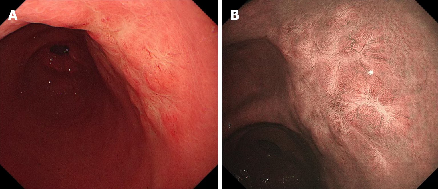
Figure 1 Endoscopic images prior to endoscopic submucosal dissection.
A: Gastroscopy revealed a mucosal rough area measuring 2.5 cm × 3.0 cm in the posterior wall of the antrum; B: Narrow band imaging showed irregular marginal crypt epithelium and subepithelial capillary network.
- Citation: Li SH, Niu WW, Huo XX, Zhang H, Shi LM, Liu YT, Xing JJ, Feng ZX, Wang N. Value of endoscopic submucosal dissection in diagnosing gastric mucosa-associated lymphoid tissue lymphoma: A case report. World J Gastroenterol 2025; 31(38): 111549
- URL: https://www.wjgnet.com/1007-9327/full/v31/i38/111549.htm
- DOI: https://dx.doi.org/10.3748/wjg.v31.i38.111549