
















©The Author(s) 2025.
World J Gastroenterol. Oct 14, 2025; 31(38): 111363
Published online Oct 14, 2025. doi: 10.3748/wjg.v31.i38.111363
Published online Oct 14, 2025. doi: 10.3748/wjg.v31.i38.111363
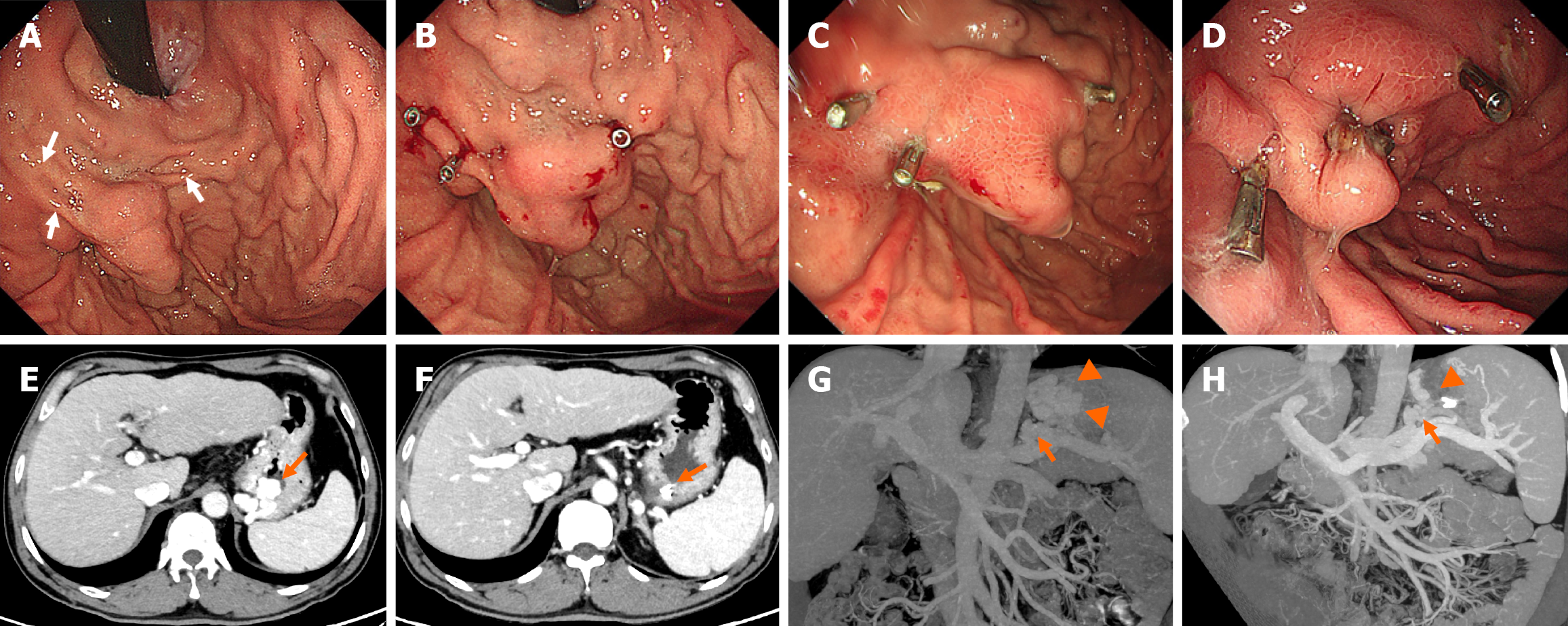
Figure 2 A 48-year-old man with tortuous gastric varices who underwent clip-assisted endoscopic cyanoacrylate injection.
A: Endoscopic examination revealed nodular gastric varices (GVs). Clips were planned to be deployed at three points (white arrows); B: Three clips were deployed on the GVs, resulting in slowing down or completely blocking the GV blood flow; C: Endoscopy revealed resolution of the GVs (with satisfactory hardening of the varix on the palpation) 30 days after the procedure; D: Endoscopy revealed postinjection ulcers 5 months after the procedure; E: Contrast-enhanced computed tomography (CT) scan showing GVs (orange arrow); F: Contrast-enhanced CT review revealed that the GVs had disappeared; the metal clips (orange arrow) could still be seen 1 week later; G: CT angiography revealed GVs (orange arrowhead) and a gastrorenal shunt (orange arrow) before the procedure; H: CT angiography revealed that the GVs had disappeared (orange arrowhead); the gastrorenal shunt was still present (orange arrow).
- Citation: Xiong YY, Li DW, Zhou TY, Ma H, Gao JG, Shen Z, Xu CF, Yu CH. Clip-assisted endoscopic cyanoacrylate injection vs endoscopic ultrasound-guided coil and cyanoacrylate injection for gastric varices: A propensity score-matched study. World J Gastroenterol 2025; 31(38): 111363
- URL: https://www.wjgnet.com/1007-9327/full/v31/i38/111363.htm
- DOI: https://dx.doi.org/10.3748/wjg.v31.i38.111363