
















©The Author(s) 2025.
World J Gastroenterol. Oct 14, 2025; 31(38): 110645
Published online Oct 14, 2025. doi: 10.3748/wjg.v31.i38.110645
Published online Oct 14, 2025. doi: 10.3748/wjg.v31.i38.110645
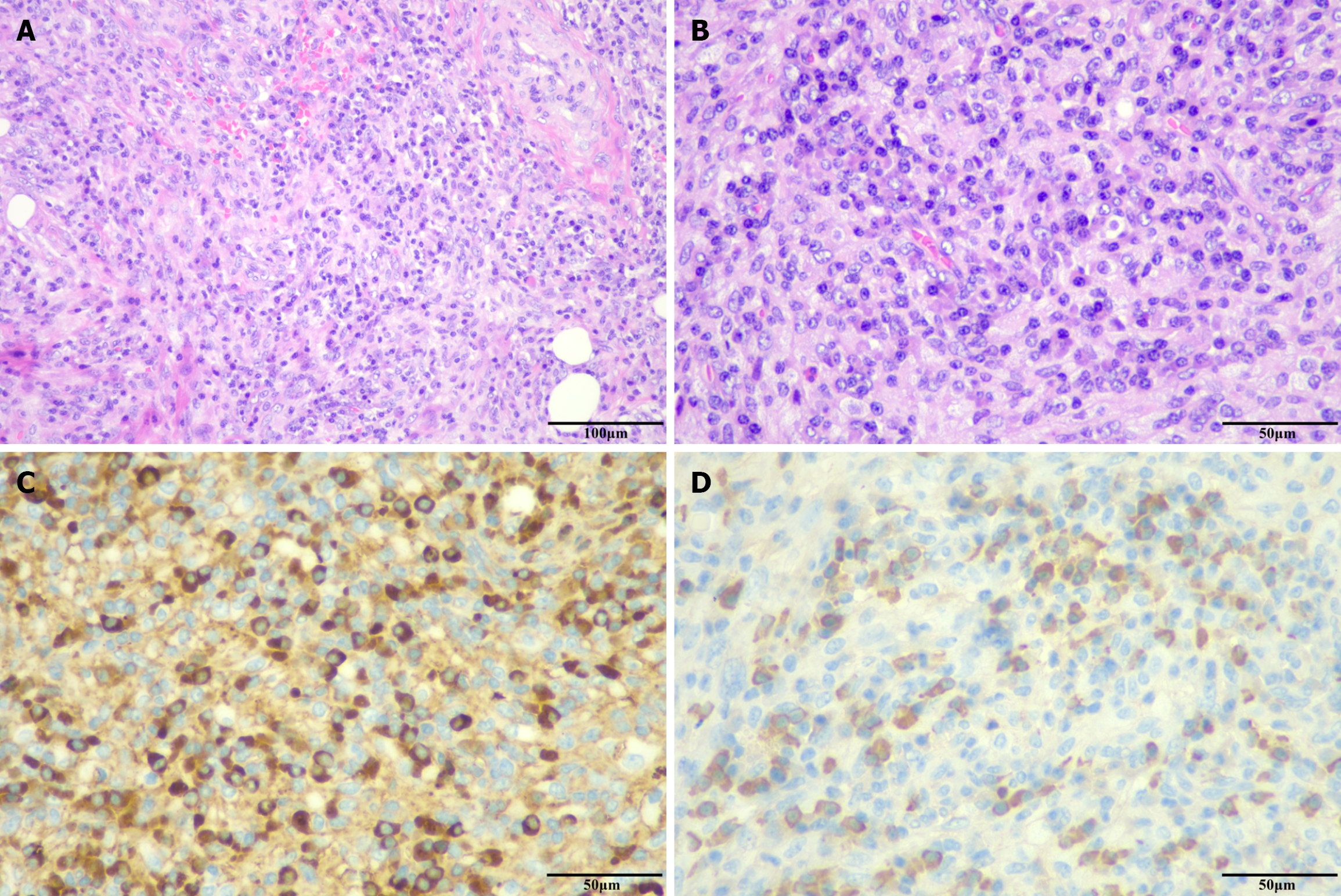
Figure 2 Pathological findings of immunoglobulin G4-related cholecystitis.
A and B: Hematoxylin-eosin staining revealed extensive infiltration of inflammatory and plasma cells, accompanied by fibrotic changes and evident phlebitis. No malignant tumor cells were observed (A: × 200; B: × 400); C: Immunoglobulin G4 (IgG4)-positive plasma cells were detected (× 400); D: IgG4-positive plasma cells were identified (× 400); they comprised more than 40% of IgG-positive plasma cells and exceeded 10 per high-power field.
- Citation: Yin ML, Ma GD, Gan YQ, Li P, Zhang ZD, Zhang SB, Meng Q, Liu B, Zhao SY. Immunoglobulin G4-related disease requiring clinical attention: A case report and review of literature. World J Gastroenterol 2025; 31(38): 110645
- URL: https://www.wjgnet.com/1007-9327/full/v31/i38/110645.htm
- DOI: https://dx.doi.org/10.3748/wjg.v31.i38.110645