
















©The Author(s) 2025.
World J Gastroenterol. Oct 14, 2025; 31(38): 110645
Published online Oct 14, 2025. doi: 10.3748/wjg.v31.i38.110645
Published online Oct 14, 2025. doi: 10.3748/wjg.v31.i38.110645
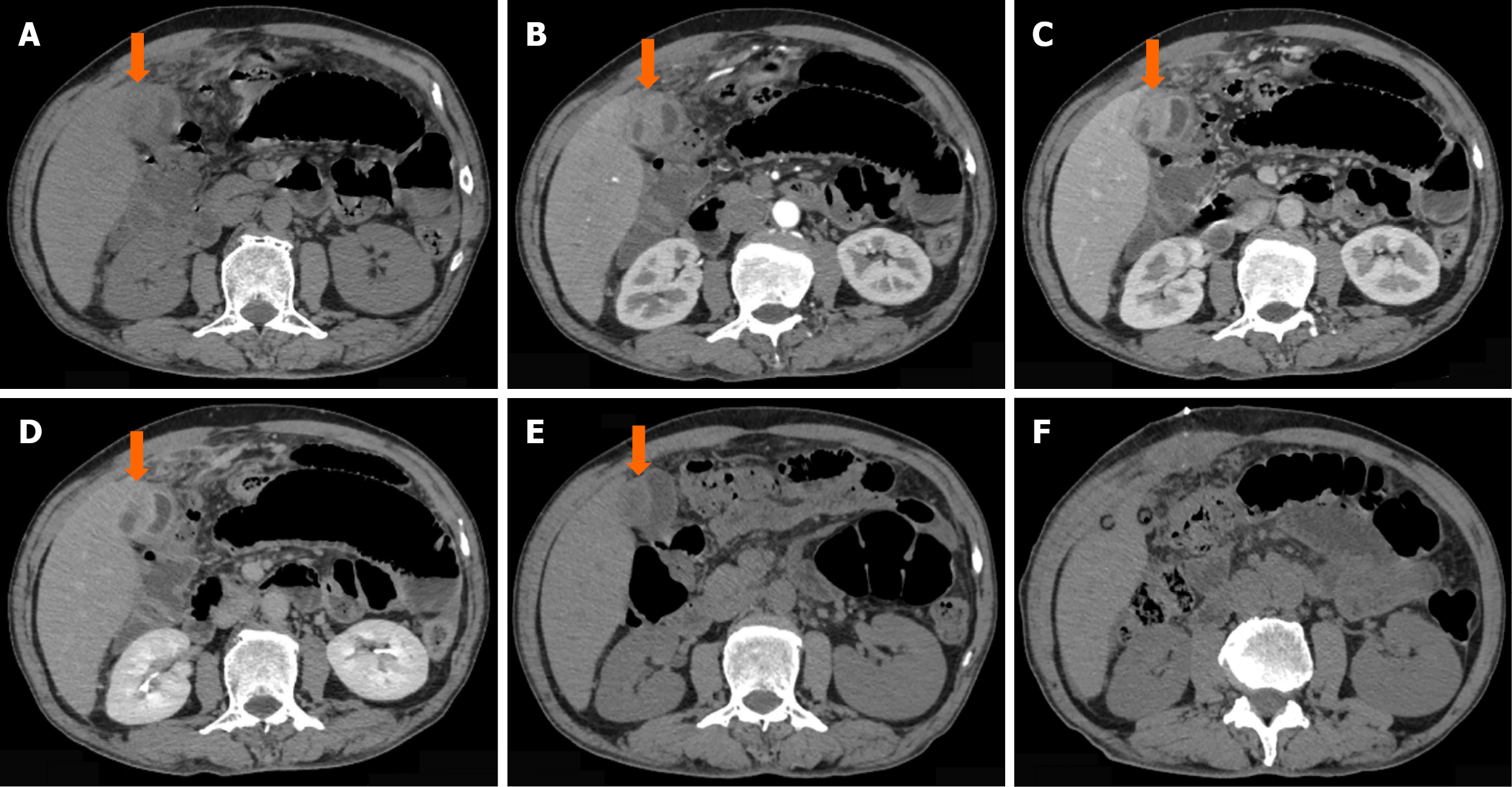
Figure 1 Imaging findings (computed tomography) of immunoglobulin G4-related cholecystitis.
A: Non-contrast computed tomography (CT) scan at the initial visit showed localized thickening at the gallbladder fundus (arrow); B: Arterial-phase contrast-enhanced CT demonstrated enhancement at the site of thickening in the gallbladder fundus (arrow); C: Venous-phase contrast-enhanced CT revealed persistent enhancement at the thickened area of the gallbladder fundus (arrow); D: Delayed-phase contrast-enhanced CT showed continued enhancement at the thickened site of the gallbladder fundus (arrow); E: Preoperative non-contrast CT scan indicated persistent localized thickening at the gallbladder fundus (arrow); F: Postoperative CT scan 1 week after surgery demonstrated good recovery in the surgical area.
- Citation: Yin ML, Ma GD, Gan YQ, Li P, Zhang ZD, Zhang SB, Meng Q, Liu B, Zhao SY. Immunoglobulin G4-related disease requiring clinical attention: A case report and review of literature. World J Gastroenterol 2025; 31(38): 110645
- URL: https://www.wjgnet.com/1007-9327/full/v31/i38/110645.htm
- DOI: https://dx.doi.org/10.3748/wjg.v31.i38.110645