
















©The Author(s) 2025.
World J Gastroenterol. Oct 14, 2025; 31(38): 110557
Published online Oct 14, 2025. doi: 10.3748/wjg.v31.i38.110557
Published online Oct 14, 2025. doi: 10.3748/wjg.v31.i38.110557
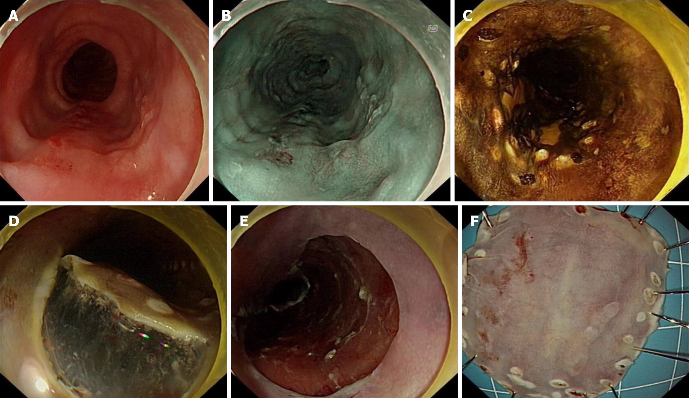
Figure 1 Endoscopic submucosal dissection of an early esophageal squamous cell carcinoma.
A: White light endoscopy to describe lesion; B: Virtual pigment description of the lesion; C: Pericyclic marker lesion; D: Submucosal dissection; E: Post-endoscopic resection of ulcer; F: Fixed specimen.
- Citation: Zhang YM, Zhu N, Chen MY, Li FL, Qin B, Jiang J, Wang SH, Wu J, Quan XJ, Wang CY, Zheng Y, Zou BC. Clinical features of early esophageal neoplastic lesions at different stages and efficacy and prognosis after endoscopic submucosal dissection. World J Gastroenterol 2025; 31(38): 110557
- URL: https://www.wjgnet.com/1007-9327/full/v31/i38/110557.htm
- DOI: https://dx.doi.org/10.3748/wjg.v31.i38.110557