
















©The Author(s) 2025.
World J Gastroenterol. Oct 14, 2025; 31(38): 109802
Published online Oct 14, 2025. doi: 10.3748/wjg.v31.i38.109802
Published online Oct 14, 2025. doi: 10.3748/wjg.v31.i38.109802
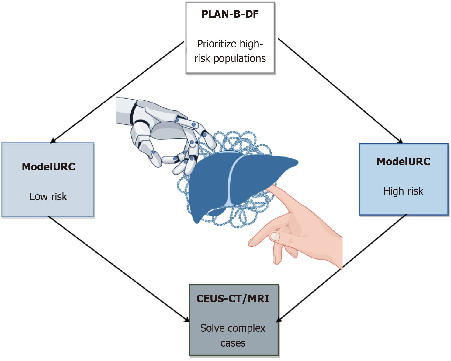
Figure 2 Integration of artificial intelligence-assisted diagnostic models and imaging techniques for liver disease evaluation.
This flowchart illustrates the risk stratification and multimodal imaging workflow for liver disease diagnosis. The PLAN-B-DF model (gradient boosting algorithm) identifies high-risk populations of hepatocellular carcinoma (HCC) in chronic hepatitis B patients by analyzing computed tomography (CT)-derived biomarkers (e.g., visceral fat distribution, spleen volume) and clinical data, achieving a C-index of 0.91. ModelURC stratifies patients into low-risk and high-risk groups for differential surveillance protocols. For complex cases (e.g., sub-3 cm lesions or atypical imaging features), contrast-enhanced ultrasound combined with CT/magnetic resonance imaging LI-RADS classification enhances diagnostic sensitivity to 88.8%, minimizing unnecessary biopsies. The framework integrates artificial intelligence-driven quantitative analysis (e.g., deep learning-based fibrosis staging, radiomics) with multimodal imaging to optimize precision in liver fibrosis grading, HCC screening, and longitudinal monitoring. CEUS: Contrast-enhanced ultrasound; CT: Computed tomography; MRI: Magnetic resonance imaging.
- Citation: Wu YM, Tang FY, Qi ZX. Multimodal artificial intelligence technology in the precision diagnosis and treatment of gastroenterology and hepatology: Innovative applications and challenges. World J Gastroenterol 2025; 31(38): 109802
- URL: https://www.wjgnet.com/1007-9327/full/v31/i38/109802.htm
- DOI: https://dx.doi.org/10.3748/wjg.v31.i38.109802