
















©The Author(s) 2025.
World J Gastroenterol. Oct 7, 2025; 31(37): 111081
Published online Oct 7, 2025. doi: 10.3748/wjg.v31.i37.111081
Published online Oct 7, 2025. doi: 10.3748/wjg.v31.i37.111081
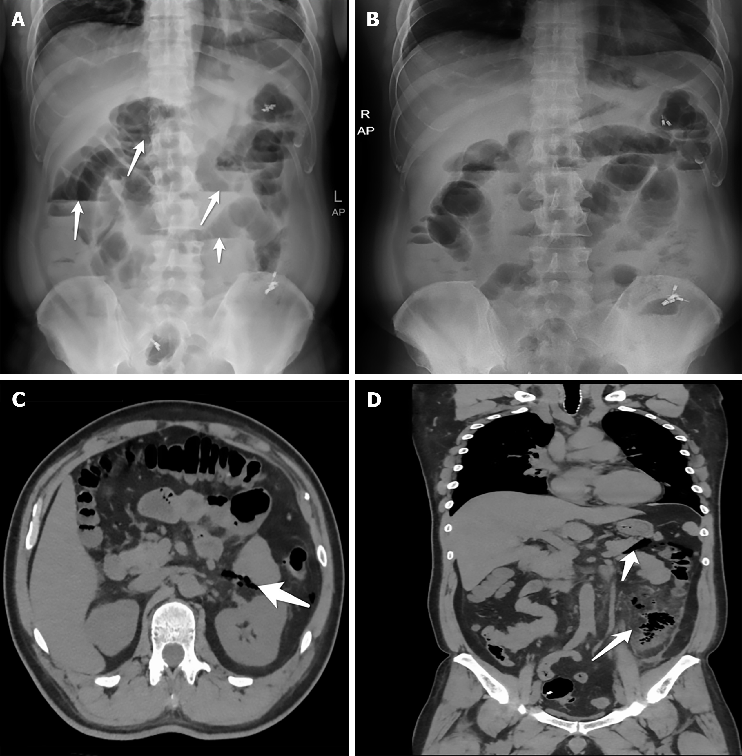
Figure 2 Initial and post treatment abdominal plain film and computed tomography scan.
A: The dilated intestinal tubes and air-fluid levels as the arrows shown after endoscopic submucosal dissection; B: Improvement of obstruction after treatment; C and D: After treatment and clip closure, computed tomography images demonstrate further the improvement of obstruction and confirm the etiology. The arrows indicated gas accumulation in the descending colon and surrounding exudation.
- Citation: Zhang Q, Hong D, Zhou YC, He GX, Jiang T. Acute intestinal obstruction caused by endoscopic submucosal dissection: A case report. World J Gastroenterol 2025; 31(37): 111081
- URL: https://www.wjgnet.com/1007-9327/full/v31/i37/111081.htm
- DOI: https://dx.doi.org/10.3748/wjg.v31.i37.111081