
















©The Author(s) 2025.
World J Gastroenterol. Sep 28, 2025; 31(36): 111137
Published online Sep 28, 2025. doi: 10.3748/wjg.v31.i36.111137
Published online Sep 28, 2025. doi: 10.3748/wjg.v31.i36.111137
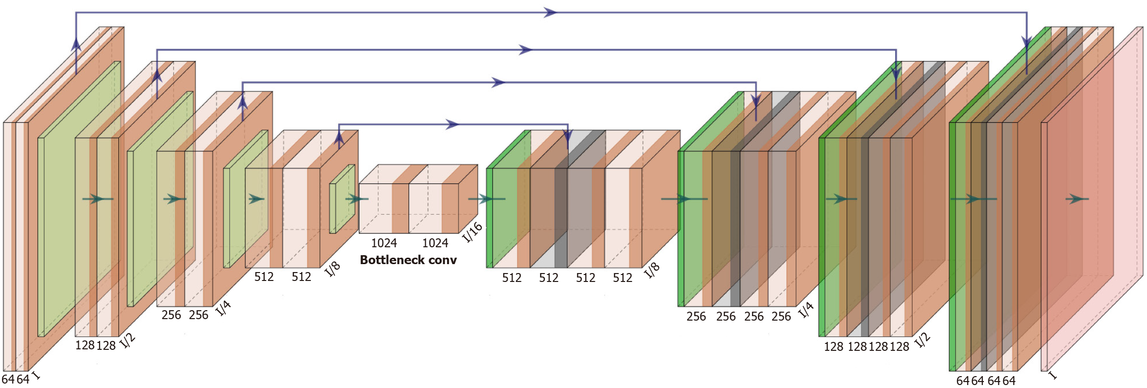
Figure 5 The U-Net architecture, widely used in medical image segmentation.
The left side is the contracting path (encoder), which reduces spatial resolution while increasing feature complexity. The right side is the expansive path (decoder), which restores spatial resolution. Skip connections link corresponding encoder-decoder levels to retain spatial detail and improve segmentation accuracy.
- Citation: Wang YY, Liu B, Wang JH. Application of deep learning-based convolutional neural networks in gastrointestinal disease endoscopic examination. World J Gastroenterol 2025; 31(36): 111137
- URL: https://www.wjgnet.com/1007-9327/full/v31/i36/111137.htm
- DOI: https://dx.doi.org/10.3748/wjg.v31.i36.111137