
















©The Author(s) 2025.
World J Gastroenterol. Sep 21, 2025; 31(35): 108139
Published online Sep 21, 2025. doi: 10.3748/wjg.v31.i35.108139
Published online Sep 21, 2025. doi: 10.3748/wjg.v31.i35.108139
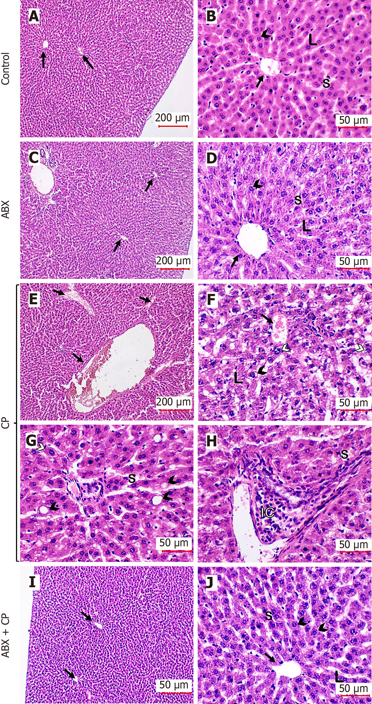
Figure 2 Ambroxol prevented liver tissue injury in cyclophosphamide-administered rats.
A-D: Photomicrographs of hematoxylin & eosin-stained sections from the control (A and B) and ABX-supplemented rats (C and D) showing normal liver architecture, including hepatocytes (arrowheads), hepatic lamina (L), central veins (arrows), and sinusoids (S); E-H: Cyclophosphamide-treated rats showing congested vessels (arrows), hepatocyte vacuolation (black arrowheads), enlarged hepatocytes with enlarged nuclei (white arrowheads), inflammatory cells (IC) infiltration, dilated sinusoids (S), and disorganized hepatic laminae (L); and I and J: ABX treatment prevented tissue injury and hepatic tissues relatively appear as control group showing normal non-congested vessels (arrows), hepatocytes (arrowheads), hepatic laminae (L), and sinusoids (S). A, C, E, and I: 100 × and scale bar = 200 µm; B, D, F, G, H and J: 400 × and scale bar = 50 µm. ABX: Ambroxol; CP: Cyclophosphamide; S: Sinusoids; L: Laminae; IC: Inflammatory cells.
- Citation: Alruhaimi RS, Hassanein EHM, Alnasser SM, Ahmeda AF, Althagafy HS, Abd El-Ghafar OAM, Mahmoud AM. Ambroxol mitigates cyclophosphamide-induced liver injury by suppressing TLR-4/NF-κB signaling and oxidative stress and upregulating cytoglobin, TXNRD1 and HMGB1. World J Gastroenterol 2025; 31(35): 108139
- URL: https://www.wjgnet.com/1007-9327/full/v31/i35/108139.htm
- DOI: https://dx.doi.org/10.3748/wjg.v31.i35.108139