
















©The Author(s) 2025.
World J Gastroenterol. Sep 14, 2025; 31(34): 110611
Published online Sep 14, 2025. doi: 10.3748/wjg.v31.i34.110611
Published online Sep 14, 2025. doi: 10.3748/wjg.v31.i34.110611
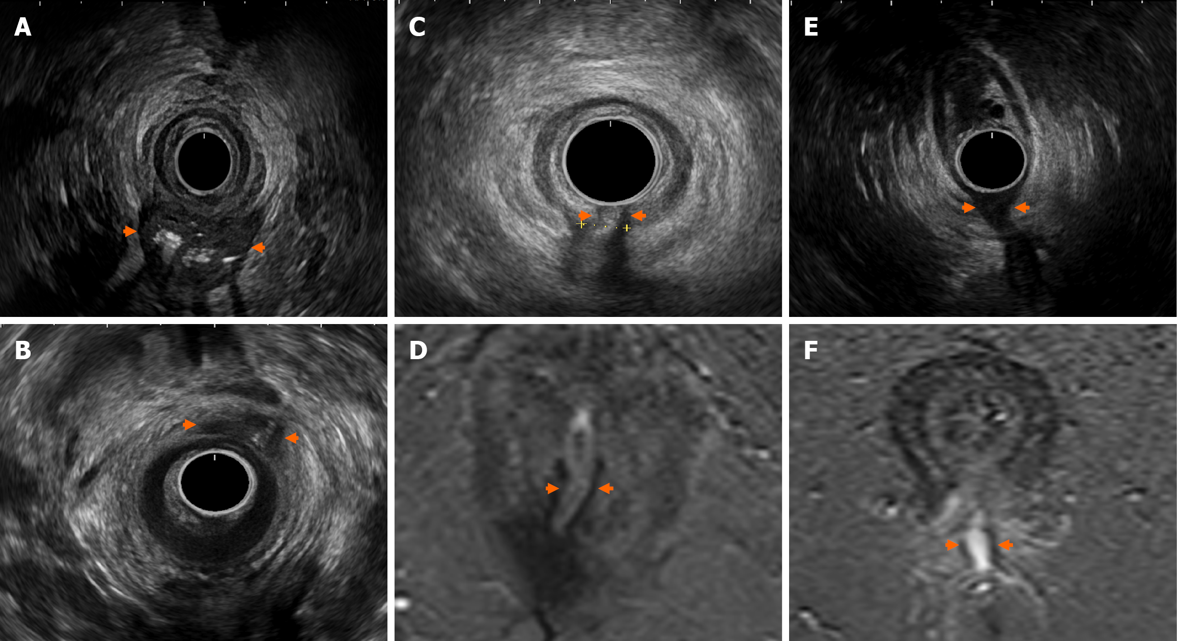
Figure 3 Evaluating perianal fistulising Crohn’s disease using endoanal ultrasound.
A: Large intersphincteric abscess with gas content (reverberation artefact from gas) posterior; B: Anterior intersphincteric abscess; C and D: Posterior midline transphincteric fistula with corresponding magnetic resonance imaging; E and F: Low transphincteric fistula with corresponding magnetic resonance imaging. Arrows delineate perianal pathology seen on endoanal ultrasound.
- Citation: Habeeb H, Chen L, De Kock I, Bhatnagar G, Kutaiba N, Vasudevan A, Srinivasan AR. Imaging in perianal fistulising Crohn’s disease: A practical guide for the gastroenterologist. World J Gastroenterol 2025; 31(34): 110611
- URL: https://www.wjgnet.com/1007-9327/full/v31/i34/110611.htm
- DOI: https://dx.doi.org/10.3748/wjg.v31.i34.110611