
















©The Author(s) 2025.
World J Gastroenterol. Sep 14, 2025; 31(34): 109900
Published online Sep 14, 2025. doi: 10.3748/wjg.v31.i34.109900
Published online Sep 14, 2025. doi: 10.3748/wjg.v31.i34.109900
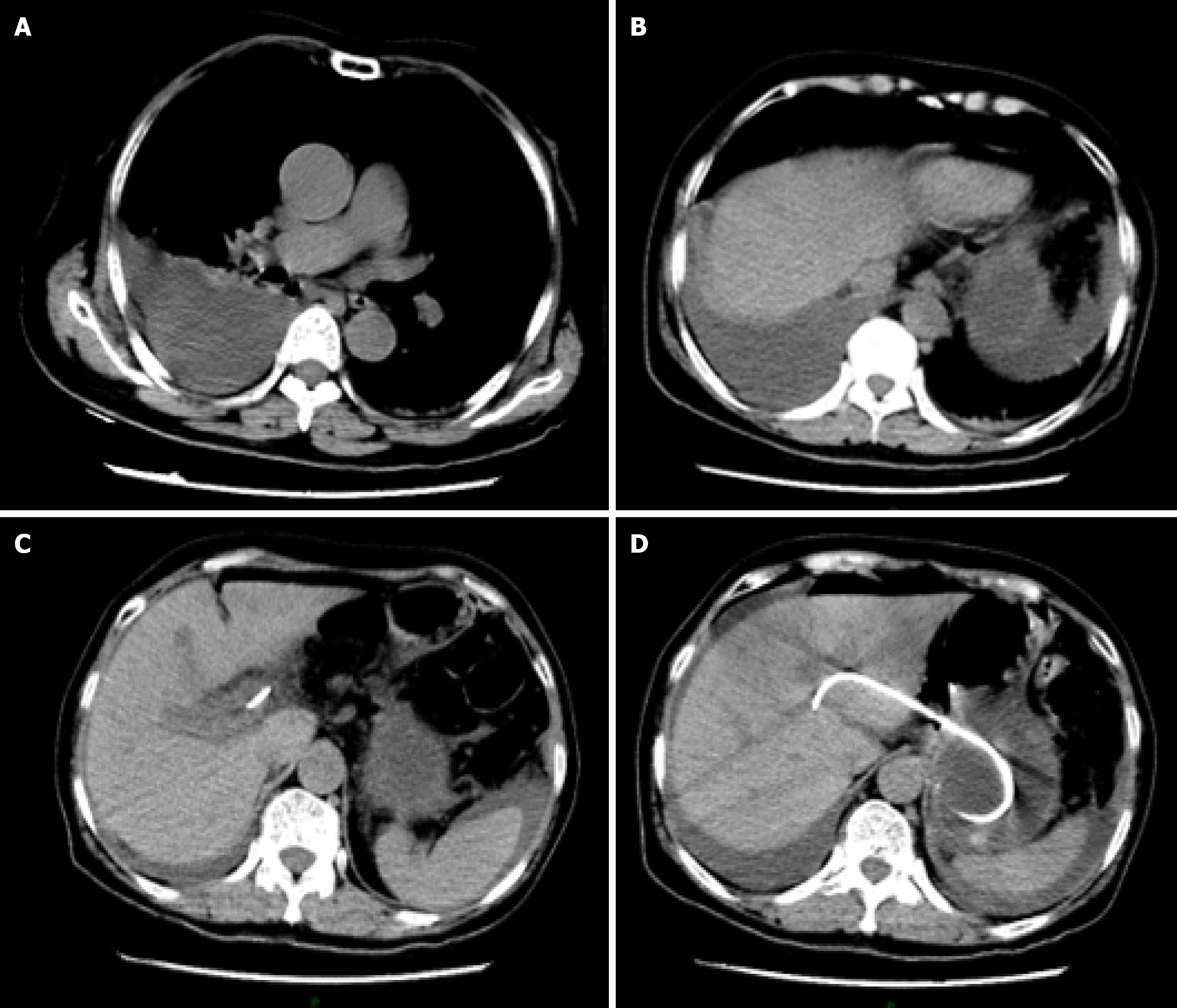
Figure 3 Emergency computed tomography scan of the patient on the second postoperative day.
Computed tomography results suggested right pleural effusion and right lower lobe lung atelectasis. The stent remained well-positioned, excluding the possibility of stent migration. A: Chest-level axial view of the patient. Density fluid opacity is identified in the right pleural cavity, occupying part of the thoracic space, consistent with right pleural effusion. The right lung tissue, compressed by the effusion, shows reduced volume and increased density, demonstrating atelectasis, with distorted and aggregated lung markings and loss of normal lung expansion morphology; B: Upper abdominal-level view of the patient. The fluid-density opacity persists in the right pleural cavity, indicating the extent of the pleural effusion. The right lung atelectasis continues, with the lung tissue compression pattern similar to that observed in view; C: Mid abdominal-level view of the patient: The shapes, sizes, and densities of upper abdominal solid organs (e.g., liver, spleen, pancreas) are approximately normal. The hollow organs (e.g., gastrointestinal tract) show normal course and morphology, without significant dilation or abnormal density; D: A stent-like foreign body shadow is visualized within the body, it remains well-positioned, same as the location of intraoperative implantation, excluding the possibility of stent migration.
- Citation: Zhang KY, He Q, Jin Y, Liu J, Lin R, Han CQ. Dark green urine following endoscopic ultrasound-guided hepaticogastrostomy: A case report. World J Gastroenterol 2025; 31(34): 109900
- URL: https://www.wjgnet.com/1007-9327/full/v31/i34/109900.htm
- DOI: https://dx.doi.org/10.3748/wjg.v31.i34.109900