
















©The Author(s) 2025.
World J Gastroenterol. Sep 14, 2025; 31(34): 109900
Published online Sep 14, 2025. doi: 10.3748/wjg.v31.i34.109900
Published online Sep 14, 2025. doi: 10.3748/wjg.v31.i34.109900
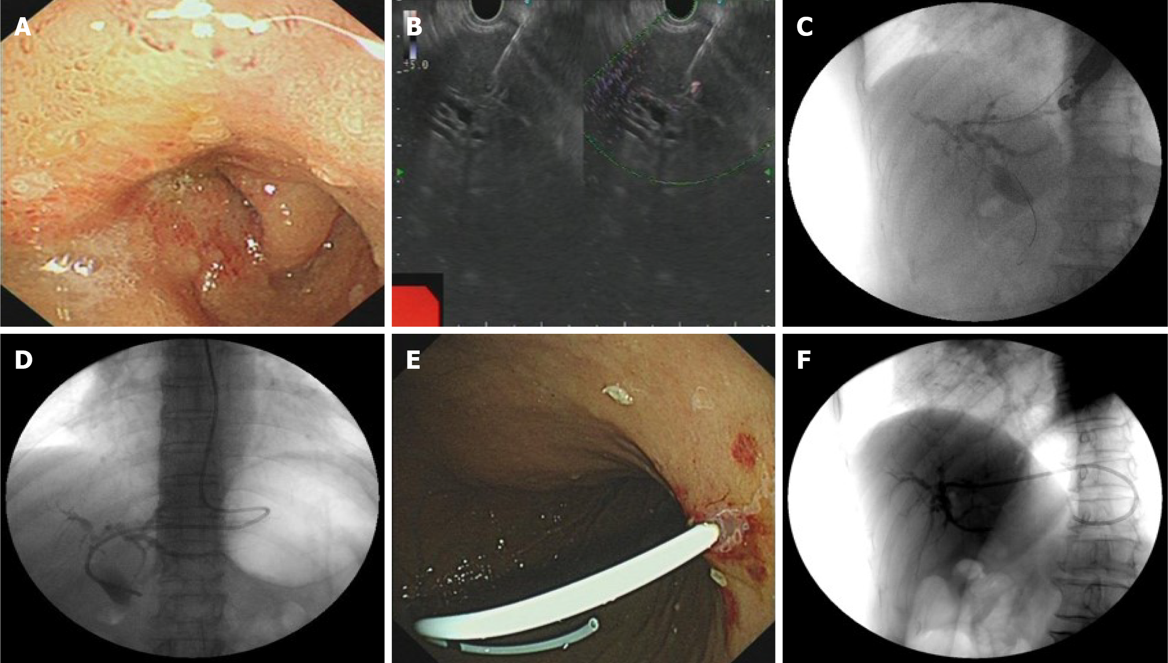
Figure 2 The Endoscopic ultrasound-guided hepaticogastrostomy process and the stent position after the operation.
A: Duodenal obstruction in the patient. Endoscopic view showing stenosis in the descending duodenum, precluding endoscopic retrograde cholangiopancreatography cannulation; B: Transgastric needle puncture. Ultrasonographic endoscopic puncture of the intrahepatic S2 segmental bile duct through the upper posterior wall of the gastric body under the cardia; C: Contrast injection. Successful injection of contrast agent (30% biligrafin) confirmings intrahepatic ductal access; D: Nasobiliary drainage placement; E: Snipped nasal bile duct for stent drainage; F: Final stent position under X-ray.
- Citation: Zhang KY, He Q, Jin Y, Liu J, Lin R, Han CQ. Dark green urine following endoscopic ultrasound-guided hepaticogastrostomy: A case report. World J Gastroenterol 2025; 31(34): 109900
- URL: https://www.wjgnet.com/1007-9327/full/v31/i34/109900.htm
- DOI: https://dx.doi.org/10.3748/wjg.v31.i34.109900