
















©The Author(s) 2025.
World J Gastroenterol. Sep 14, 2025; 31(34): 109825
Published online Sep 14, 2025. doi: 10.3748/wjg.v31.i34.109825
Published online Sep 14, 2025. doi: 10.3748/wjg.v31.i34.109825
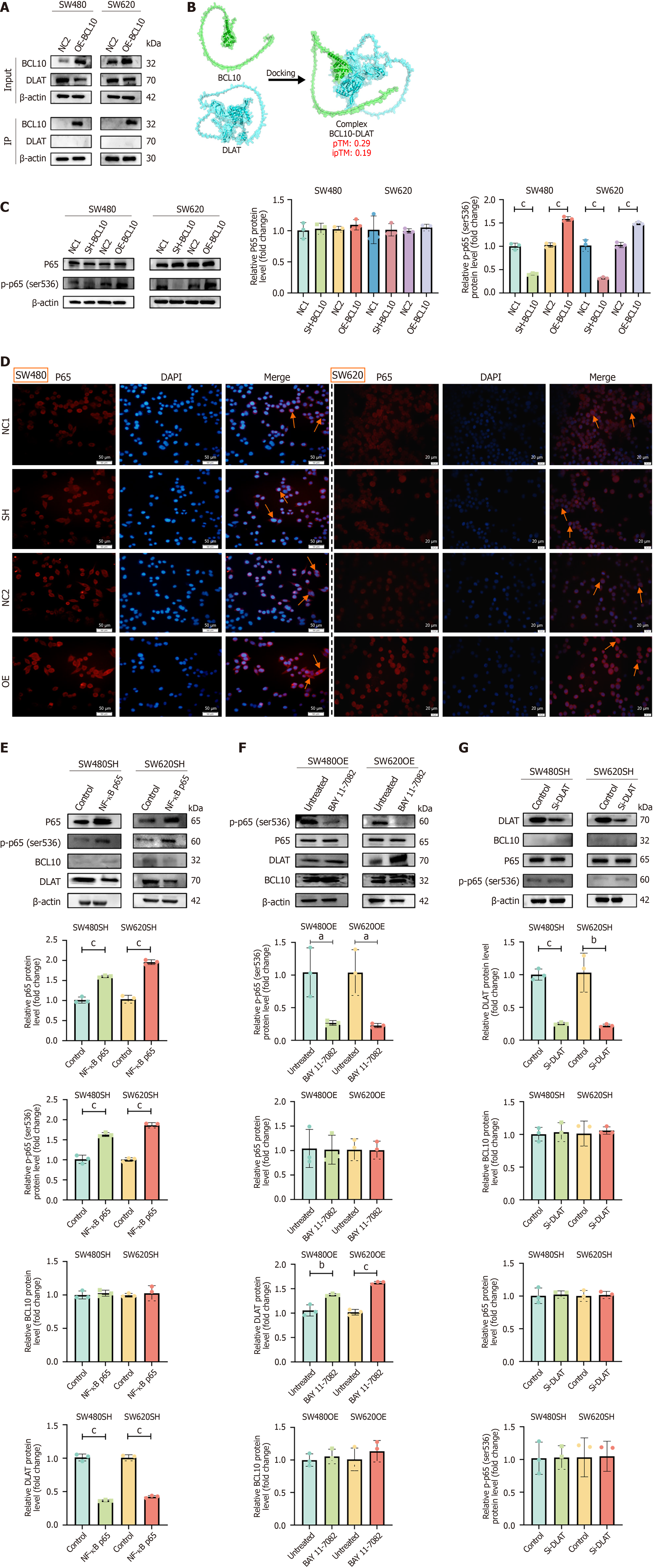
Figure 5 B-cell CLL/Lymphoma 10 regulates DLAT through NF-κB p65 phosphorylation.
A: Co-IP of DLAT and B-cell CLL/Lymphoma 10 (BCL10); B: AlphaFold3 predicted binding model (ipTM < 0.6); C: Western blot of p65 and p-p65 (Ser536). cP < 0.001; D: Immunofluorescence of p65 (red) and DAPI (blue); orange arrows indicate nuclear translocation; E: Western blot after p65 transfection in BCL10-knockdown cells. cP < 0.001; F: Western blot after BAY 11-7082 treatment. aP < 0.05. bP < 0.01. cP < 0.001; G: Western blot after DLAT knockout. cP < 0.001.
- Citation: Xiao PT, Li CF, Liu YD, Zhong J, Cui XL, Liu C, Yang W. B cell CLL/lymphoma 10 promotes colorectal cancer cell proliferation and regulates cuproptosis sensitivity through the NF-κB signaling pathway. World J Gastroenterol 2025; 31(34): 109825
- URL: https://www.wjgnet.com/1007-9327/full/v31/i34/109825.htm
- DOI: https://dx.doi.org/10.3748/wjg.v31.i34.109825