
















©The Author(s) 2025.
World J Gastroenterol. Sep 14, 2025; 31(34): 108807
Published online Sep 14, 2025. doi: 10.3748/wjg.v31.i34.108807
Published online Sep 14, 2025. doi: 10.3748/wjg.v31.i34.108807
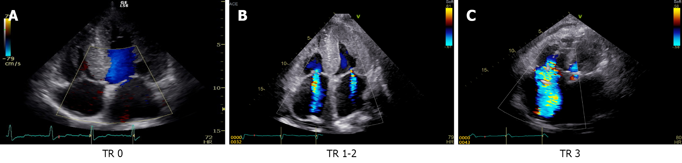
Figure 2 Transthoracic echocardiographic images demonstrating three grades of tricuspid regurgitation severity using color Doppler flow mapping.
A: Mild tricuspid regurgitation (TR): A narrow, faint regurgitant jet confined to the tricuspid valve plane; B: Moderate TR: A more prominent jet extending into the right atrium but not reaching the posterior wall; C: Severe TR: A broad, dense jet filling more than half of the right atrium with systolic flow reversal in the hepatic veins. TR: Tricuspid regurgitation.
- Citation: Miida S, Kamimura H, Fujiki S, Kobayashi T, Endo S, Maruyama H, Yoshida T, Watanabe Y, Kimura N, Abe H, Sakamaki A, Yokoo T, Tsukada M, Numano F, Kashimura T, Inomata T, Fuzawa Y, Hirata T, Horii Y, Ishikawa H, Nonaka H, Kamimura K, Terai S. Image analysis of cardiac hepatopathy secondary to heart failure: Machine learning vs gastroenterologists and radiologists. World J Gastroenterol 2025; 31(34): 108807
- URL: https://www.wjgnet.com/1007-9327/full/v31/i34/108807.htm
- DOI: https://dx.doi.org/10.3748/wjg.v31.i34.108807