
















©The Author(s) 2025.
World J Gastroenterol. Sep 14, 2025; 31(34): 108807
Published online Sep 14, 2025. doi: 10.3748/wjg.v31.i34.108807
Published online Sep 14, 2025. doi: 10.3748/wjg.v31.i34.108807
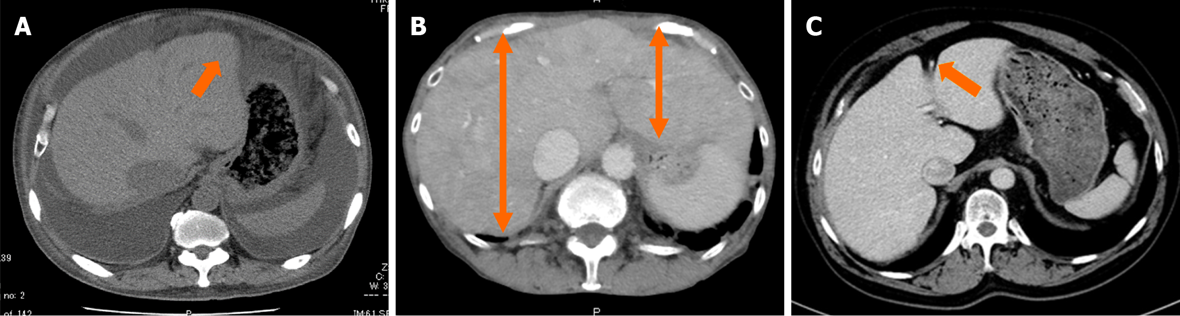
Figure 1 Imaging features of congestive liver.
A: Ascites and slightly blunted liver edges in severe cases (orange arrow); B: Enlargement of the hepatic veins and Right and Left lobe enlargement (orange bidirectional arrow); C: Images at the paraumbilical vein, dilation of the paraumbilical vein, a component of the portal venous system, is observed (orange arrow).
- Citation: Miida S, Kamimura H, Fujiki S, Kobayashi T, Endo S, Maruyama H, Yoshida T, Watanabe Y, Kimura N, Abe H, Sakamaki A, Yokoo T, Tsukada M, Numano F, Kashimura T, Inomata T, Fuzawa Y, Hirata T, Horii Y, Ishikawa H, Nonaka H, Kamimura K, Terai S. Image analysis of cardiac hepatopathy secondary to heart failure: Machine learning vs gastroenterologists and radiologists. World J Gastroenterol 2025; 31(34): 108807
- URL: https://www.wjgnet.com/1007-9327/full/v31/i34/108807.htm
- DOI: https://dx.doi.org/10.3748/wjg.v31.i34.108807