
















©The Author(s) 2025.
World J Gastroenterol. Sep 14, 2025; 31(34): 108623
Published online Sep 14, 2025. doi: 10.3748/wjg.v31.i34.108623
Published online Sep 14, 2025. doi: 10.3748/wjg.v31.i34.108623
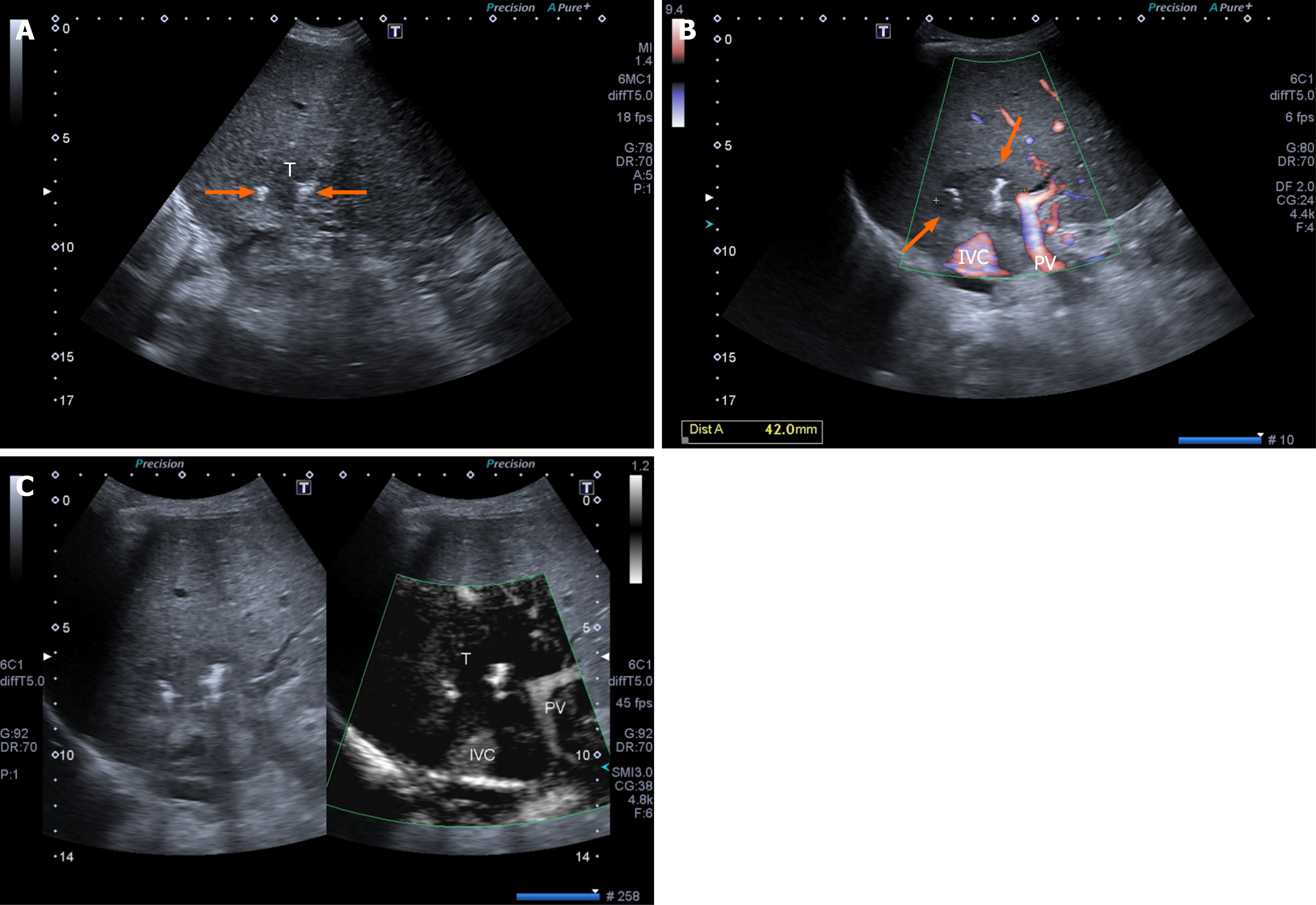
Figure 2 Irreversible electroporation.
A: A 3 cm intrahepatic cholangiocarcinoma hypoechoic nodule (arrows) is located between the inferior vena cava and the main portal trunk as shown by power Doppler ultrasound (US); B: US appearance of two probes (arrows) inserted into the tumor; C: Contrast-enhanced US shows complete necrosis of the tumor. IVC: Inferior vena cava; PV: Portal vein; T: Intrahepatic cholangiocarcinoma nodule.
- Citation: Giorgio A, Ciracì E, De Luca M, Stella G, Rollo VC, Montesarchio L, Giorgio V. Ultrasound-guided percutaneous thermal and non-thermal ablation of intrahepatic cholangiocarcinoma. World J Gastroenterol 2025; 31(34): 108623
- URL: https://www.wjgnet.com/1007-9327/full/v31/i34/108623.htm
- DOI: https://dx.doi.org/10.3748/wjg.v31.i34.108623