
















©The Author(s) 2025.
World J Gastroenterol. Sep 14, 2025; 31(34): 108623
Published online Sep 14, 2025. doi: 10.3748/wjg.v31.i34.108623
Published online Sep 14, 2025. doi: 10.3748/wjg.v31.i34.108623
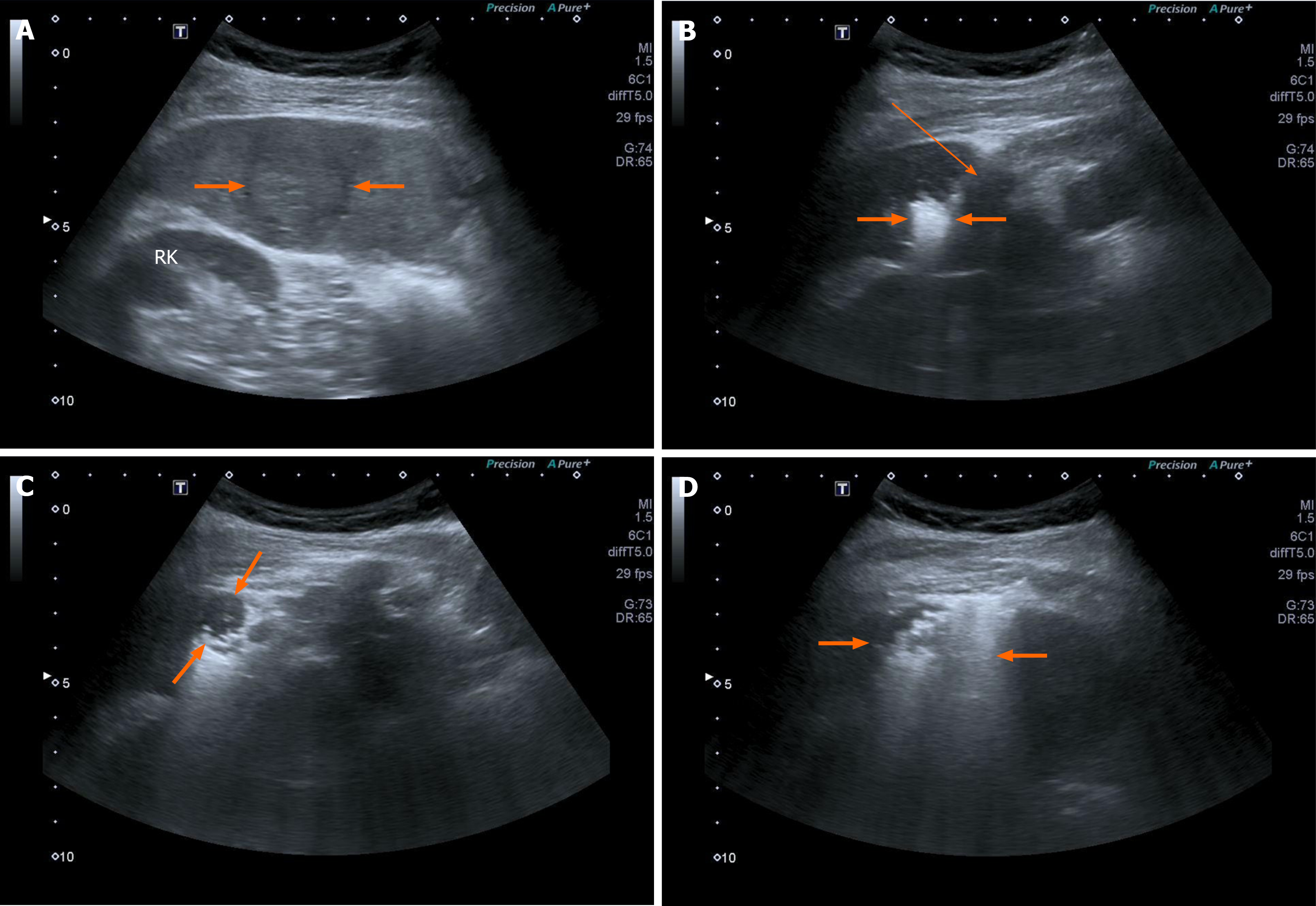
Figure 1 Microwave ablation.
A: Intrahepatic cholangiocarcinoma (arrows) in the sixth segment of the liver; B: The antenna (subtle arrow) is inserted into the tumor, and the generator is on. See the time in the top right. The hyperechoic appearance (arrows) corresponds to the steam generated by heat and reflects the ablated area from microwave application; C: After 1 minute, almost all nodules become hyperechoic except for a small, round, hypoechoic area (arrows) in the top left; D: The entire nodule is hyperechoic (arrows) after 2 minutes from the start of microwave application, indicating complete necrosis of the nodule. T: Intrahepatic cholangiocarcinoma nodule.
- Citation: Giorgio A, Ciracì E, De Luca M, Stella G, Rollo VC, Montesarchio L, Giorgio V. Ultrasound-guided percutaneous thermal and non-thermal ablation of intrahepatic cholangiocarcinoma. World J Gastroenterol 2025; 31(34): 108623
- URL: https://www.wjgnet.com/1007-9327/full/v31/i34/108623.htm
- DOI: https://dx.doi.org/10.3748/wjg.v31.i34.108623