
















©The Author(s) 2025.
World J Gastroenterol. Sep 14, 2025; 31(34): 108617
Published online Sep 14, 2025. doi: 10.3748/wjg.v31.i34.108617
Published online Sep 14, 2025. doi: 10.3748/wjg.v31.i34.108617
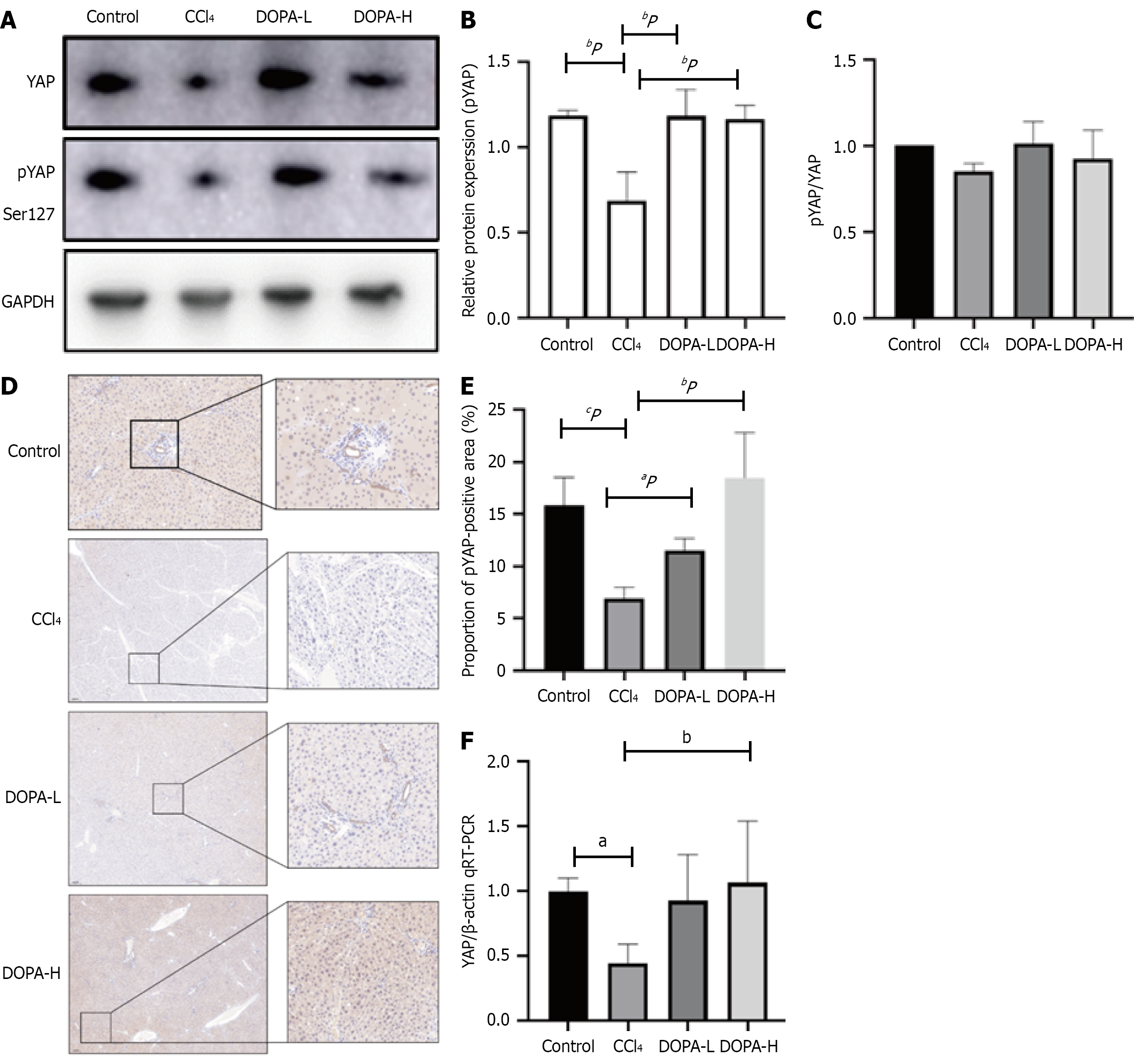
Figure 4 Phosphorylation of Yes-associated protein 1 in Hippo signaling pathway.
A: Western blotting was used to analyze the expression of p-Yes-associated protein 1 (YAP) in lysed liver tissue, and the results were standardized relative to GAPDH expression (n = 8); B: The representative protein bands were shown on the corresponding histogram; C: The ratio of pYAP to YAP is shown; D: Representative fiber images of liver pYAP immunohistochemistry (original magnification, × 100). Scale bar = 100 μm; E: ImageJ software (NIH) was used for quantitative analysis of DRD1 positive area, n = 8/group; F: Quantitative real-time polymerase chain reaction was used to detect the mRNA expression level of YAP. aP < 0.05; bP < 0.01; cP < 0.001; NS: Not significant; CCl4: Carbon tetrachloride; YAP: Yes-associated protein 1; qRT-PCR: Reverse transcription-quantitative real-time polymerase chain reaction.
- Citation: Wang HY, Qi MM, Zhang K, Zhu YZ, Zhang J. Dopamine receptor D1-mediated suppression of liver fibrosis via Hippo/Yes-associated protein 1 signaling in levodopa treatment. World J Gastroenterol 2025; 31(34): 108617
- URL: https://www.wjgnet.com/1007-9327/full/v31/i34/108617.htm
- DOI: https://dx.doi.org/10.3748/wjg.v31.i34.108617