
















©The Author(s) 2025.
World J Gastroenterol. Sep 7, 2025; 31(33): 109811
Published online Sep 7, 2025. doi: 10.3748/wjg.v31.i33.109811
Published online Sep 7, 2025. doi: 10.3748/wjg.v31.i33.109811
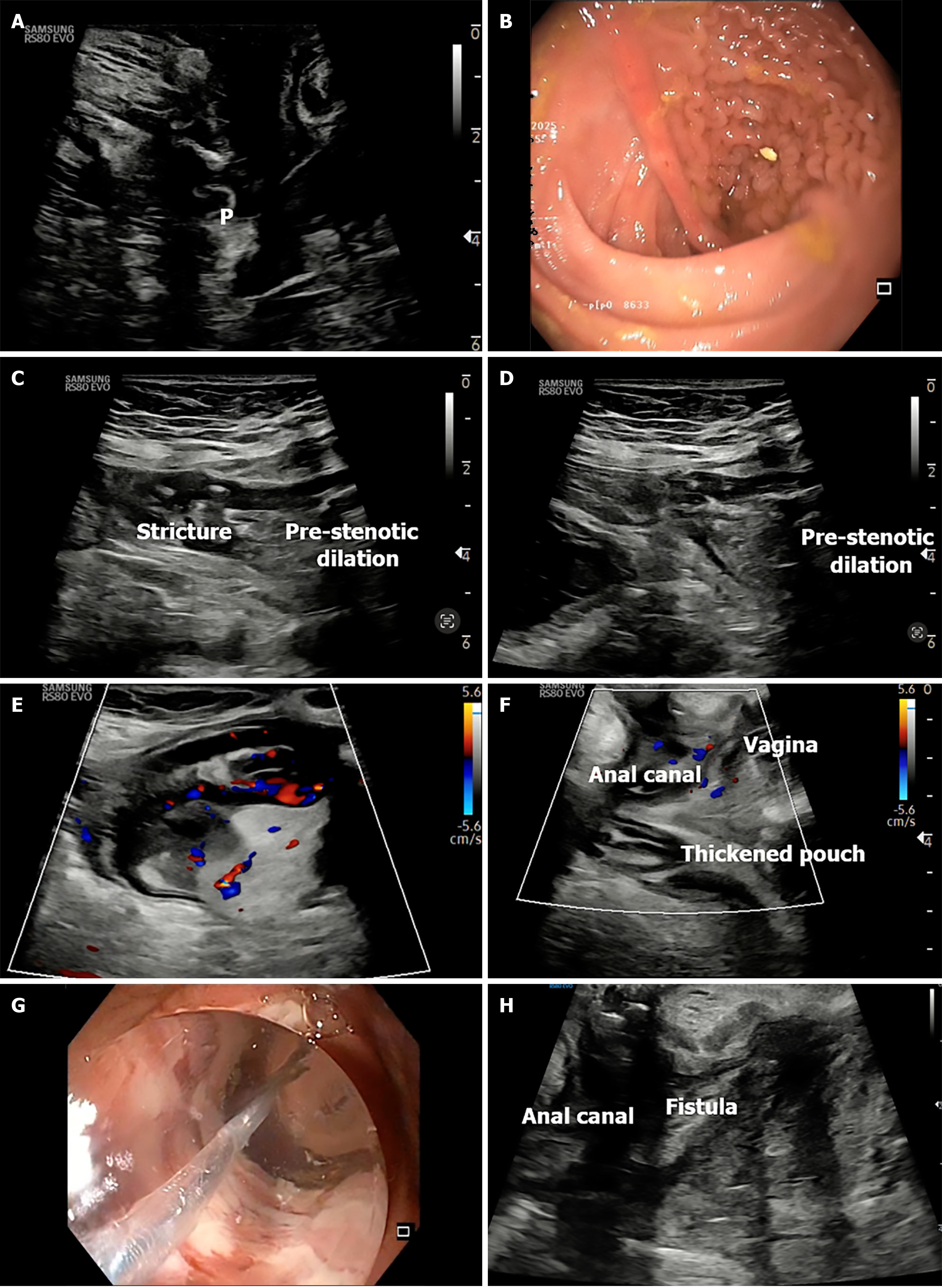
Figure 9 Transperineal ultrasound in ileal pouch.
A: Transperineal ultrasound (TPUS) image showing normal pouch wall; B: Endoscopic view of normal pouch mucosa; C and D: Transabdominal ultrasound showing prestenotic dilatation with stricture; E: Color Doppler showing increased vascularity in the pouch; F: TPUS sagittal image of thickened pouch with adjacent vagina; G: Endoscopic balloon dilation under image guidance; H: TPUS view of anorectum and vagina for assessing anovaginal fistula in in ileal pouch case. P: Pouch wall.
- Citation: Pal P, Mateen MA, Pooja K, Gupta R, Tandan M, Reddy DN. Transperineal ultrasound: Role in inflammatory bowel disease management. World J Gastroenterol 2025; 31(33): 109811
- URL: https://www.wjgnet.com/1007-9327/full/v31/i33/109811.htm
- DOI: https://dx.doi.org/10.3748/wjg.v31.i33.109811