
















©The Author(s) 2025.
World J Gastroenterol. Sep 7, 2025; 31(33): 109811
Published online Sep 7, 2025. doi: 10.3748/wjg.v31.i33.109811
Published online Sep 7, 2025. doi: 10.3748/wjg.v31.i33.109811
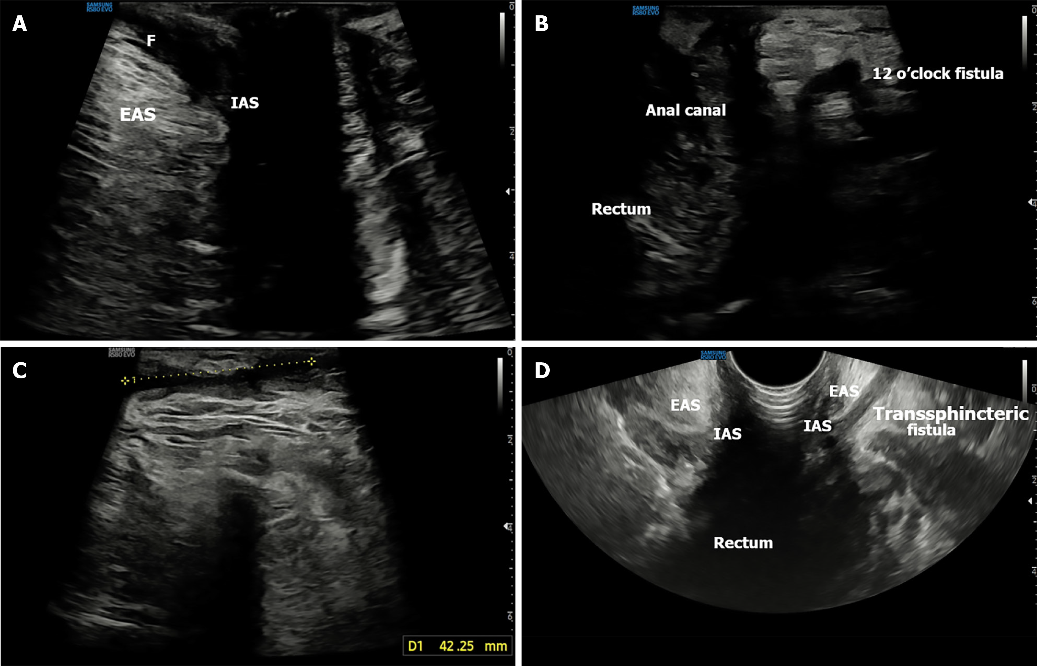
Figure 5 Inter- and trans- sphincteric fistula.
A: Transperineal ultrasound sagittal image showing intersphincteric fistula between internal anal sphncter and extenal anal sphicter (EAS); B: Transsphincteric fistula originating from anal canal and extending through EAS (12 o’clock); C: Axial view showing orientation of the long tract going anteriorly; D: Complex transsphincteric tract with anterior extension seen on transrectal ultrasound. F: Fistula; EAS: Extenal anal sphincter; IAS: Internal anal sphincter.
- Citation: Pal P, Mateen MA, Pooja K, Gupta R, Tandan M, Reddy DN. Transperineal ultrasound: Role in inflammatory bowel disease management. World J Gastroenterol 2025; 31(33): 109811
- URL: https://www.wjgnet.com/1007-9327/full/v31/i33/109811.htm
- DOI: https://dx.doi.org/10.3748/wjg.v31.i33.109811