
















©The Author(s) 2025.
World J Gastroenterol. Sep 7, 2025; 31(33): 105466
Published online Sep 7, 2025. doi: 10.3748/wjg.v31.i33.105466
Published online Sep 7, 2025. doi: 10.3748/wjg.v31.i33.105466
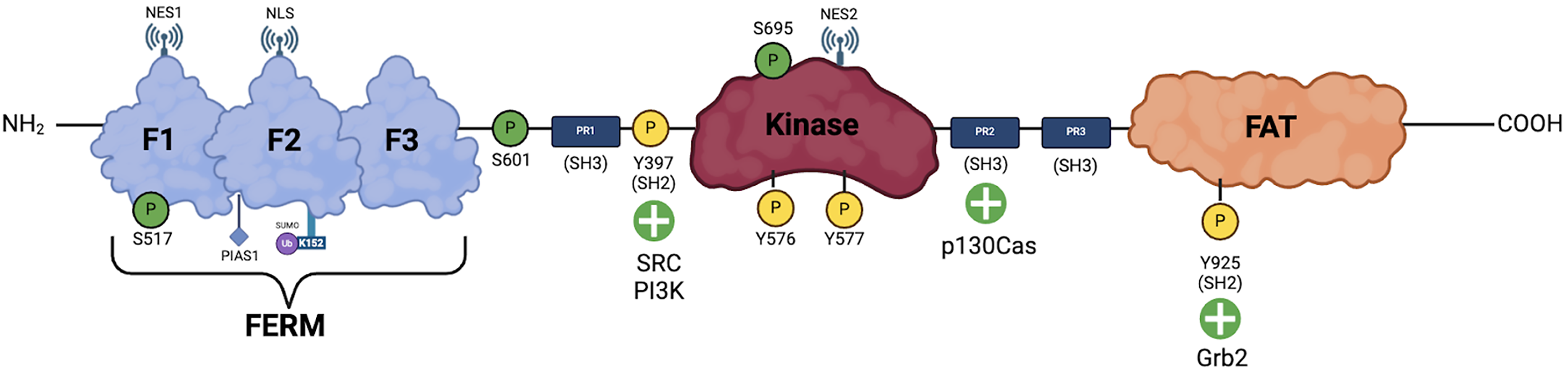
Figure 2 Focal adhesion kinase phosphorylation.
The figure illustrates the phosphorylation of focal adhesion kinase and its interactions with various proteins, highlighting the regulatory mechanisms that govern focal adhesion kinase activation and signaling. Green “P” icons represent the various serine residues that undergo protein kinase B 1 (Akt1)-mediated phosphorylation, yellow “P” icons represent the various tyrosine residues that undergo phosphorylation by various mechanisms. NES: Nuclear export signal; NLS: Nuclear localization signals; PIAS1: Protein inhibitor of activated Stat1; Ub: Ubiquitin; SUMO: Small ubiquitin-related modifier; PR: Progesterone receptor; SH: Src homology domain; PI3K: Phosphoinositide 3-kinase; FAT: Focal adhesion targeting; Grb2: Growth factor receptor-bound protein 2.
- Citation: Malek KK, Kumar B, Ahmad R, Singh A, Basson MD. Focal adhesion kinase: A promising regulator of colitis-associated healing. World J Gastroenterol 2025; 31(33): 105466
- URL: https://www.wjgnet.com/1007-9327/full/v31/i33/105466.htm
- DOI: https://dx.doi.org/10.3748/wjg.v31.i33.105466