
















©The Author(s) 2025.
World J Gastroenterol. Aug 28, 2025; 31(32): 110573
Published online Aug 28, 2025. doi: 10.3748/wjg.v31.i32.110573
Published online Aug 28, 2025. doi: 10.3748/wjg.v31.i32.110573
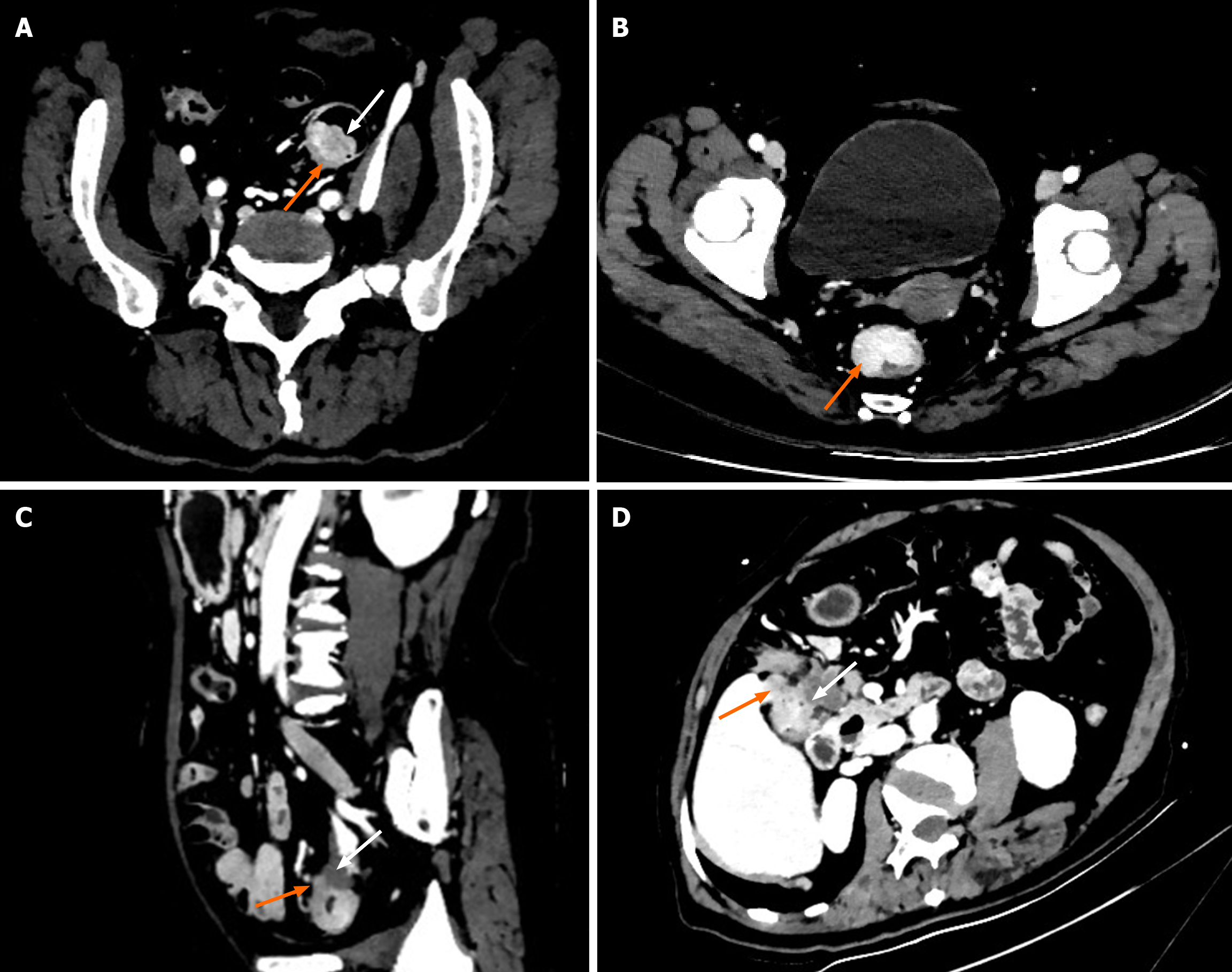
Figure 1 Computed tomography staging criteria.
A: A 73-year-old male patient with histological stage T1 tumor. The 40 keV oblique axial (OA) virtual monoenergetic image in venous phase showed a sigmoid colon mucosal mass (white arrow) surrounding 1/3 of the lumen with clearly visible normal bowel wall muscularis propria appearing (orange arrow) with relatively low density and without peritumoral fat stranding (PFS) indicative of T1 stage disease; B: A 53-year-old female patient with histological stage T2 tumor. The 40 keV OA virtual monoenergetic image in venous phase showed an upper rectal tumor (orange arrow) surrounding 2/3 of the lumen with a smooth outer border of thickened rectal wall and without PFS indicative of T2 stage disease; C: A 65-year-old male patient with histological stage T3 tumor. The 40 keV OA virtual monoenergetic image in venous phase demonstrated a sigmoid colon tumor with an irregular border and (orange arrow) spiculations extending into the peri-rectal fat, along with PFS sign (white arrow) - findings indicative of stage T3 disease; D: A 63-year-old male patient with histological stage T4 tumor. The 40 keV OA virtual monoenergetic image in venous phase demonstrated a tumor at the hepatic flexure surrounding the entire lumen (white arrow) with infiltration into peritumoral fat and invasion of adjacent liver (orange arrow) indicative of T4 stage disease.
- Citation: Chen FX, Jiang KK, Zhu JF, Wang MR, Fan XL, Yang JS, He BS. Diagnostic accuracy of dual-layer spectral computed tomography virtual monoenergetic imaging with multiplanar reformation for T-staging of colorectal cancer. World J Gastroenterol 2025; 31(32): 110573
- URL: https://www.wjgnet.com/1007-9327/full/v31/i32/110573.htm
- DOI: https://dx.doi.org/10.3748/wjg.v31.i32.110573