
















©The Author(s) 2025.
World J Gastroenterol. Aug 28, 2025; 31(32): 108654
Published online Aug 28, 2025. doi: 10.3748/wjg.v31.i32.108654
Published online Aug 28, 2025. doi: 10.3748/wjg.v31.i32.108654
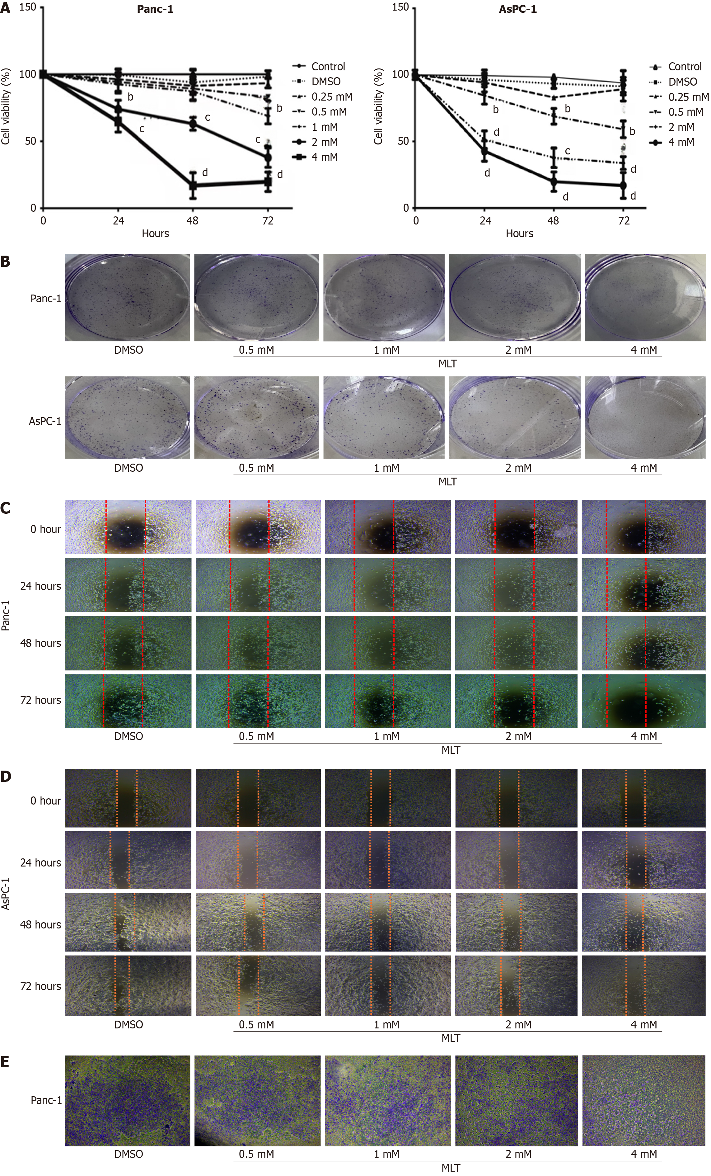
Figure 1 Effects of melatonin on cell viability, colony formation, migration, and invasion in pancreatic cancer.
A: Panc-1 and AsPC-1 cells were treated with different melatonin concentrations (0.25, 0.5, 1, 2, and 4 mmol/L) or vehicle (0.1% dimethyl sulfoxide) for 0, 24, 48, or 72 hours. Cell viability was assessed using the cell counting kit-8 assay (n = 3); B: Effects of different melatonin concentrations on colony formation in Panc-1 and AsPC-1 cells; C and D: Scratch assay showing the effects of different melatonin concentrations on the migration of Panc-1 and AsPC-1 cells; E: Invasion of Panc-1 cells treated with different melatonin concentrations for 48 hours observed at a magnification of 100 ×. All images show representative images from three independent experiments. MLT: Melatonin; DMSO: Dimethyl sulfoxide. bP < 0.01 vs control, cP < 0.001 vs control, and dP < 0.0001 vs control.
- Citation: Zhao Q, Zhang H, Wu HM, Yang QY, Zhao H, Kang L, Lv XY. Melatonin-induced ferroptosis in pancreatic cancer cells by stimulating endoplasmic reticulum stress and inhibiting alanine-serine-cysteine transporter 2-driven glutamine metabolism. World J Gastroenterol 2025; 31(32): 108654
- URL: https://www.wjgnet.com/1007-9327/full/v31/i32/108654.htm
- DOI: https://dx.doi.org/10.3748/wjg.v31.i32.108654