
















©The Author(s) 2025.
World J Gastroenterol. Aug 28, 2025; 31(32): 106424
Published online Aug 28, 2025. doi: 10.3748/wjg.v31.i32.106424
Published online Aug 28, 2025. doi: 10.3748/wjg.v31.i32.106424
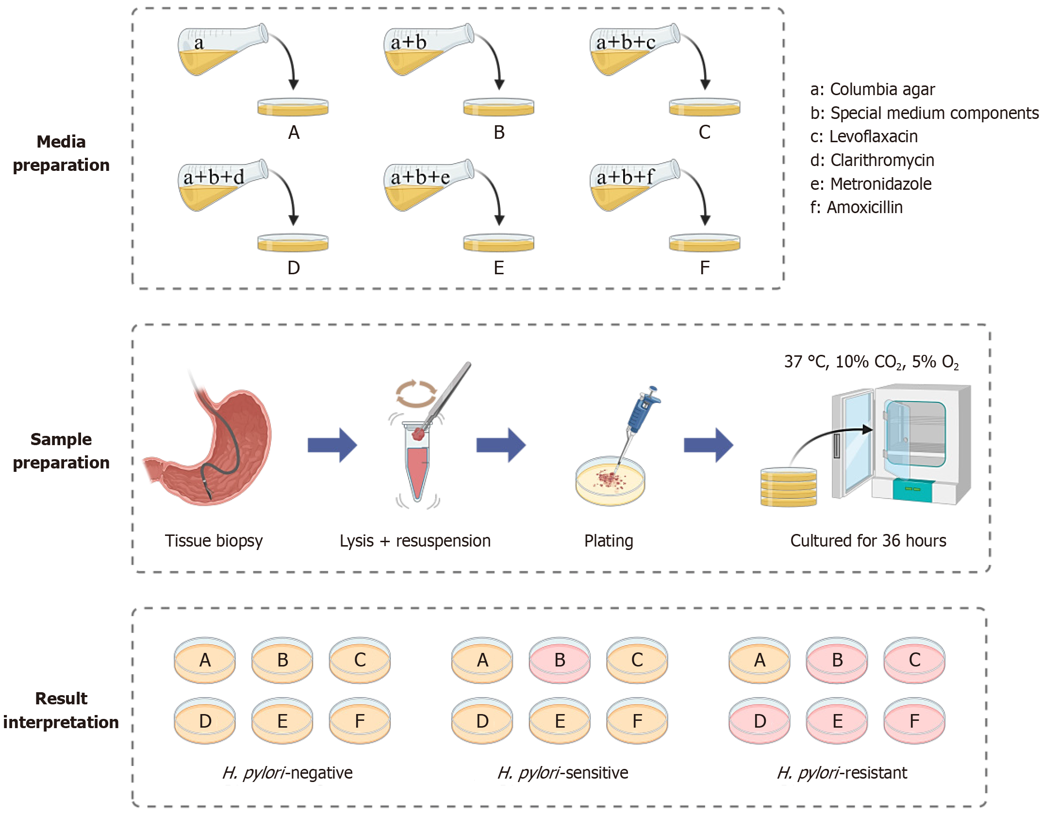
Figure 1 Schematic illustration of the rapid detection of antibiotic resistance in Helicobacter pylori.
For a given bacterial strain, its resistance to specific antibiotics is determined by the color change of the medium. The yellowish color of the medium indicates that no Helicobacter pylori growth has occurred, whereas the red color indicates the presence of Helicobacter pylori growth. In particular, the appearance of a red color in the medium was employed as an indicator of bacterial resistance. A: It represents Columbia medium; B: It stands for characteristic medium without antibiotics; C: It denotes characteristic medium with levofloxacin; D: It indicates characteristic medium with clarithromycin; E: It refers to characteristic medium with metronidazole; F: It symbolizes characteristic medium with amoxicillin. H. pylori: Helicobacter pylori.
- Citation: Guan AX, Yang SY, Wu T, Zhou WT, Chen H, Huang ZS, Luo PP, Huang YQ. Novel chromogenic medium-based method for the rapid detection of Helicobacter pylori drug resistance. World J Gastroenterol 2025; 31(32): 106424
- URL: https://www.wjgnet.com/1007-9327/full/v31/i32/106424.htm
- DOI: https://dx.doi.org/10.3748/wjg.v31.i32.106424