
















©The Author(s) 2022.
World J Gastroenterol. Oct 28, 2022; 28(40): 5827-5844
Published online Oct 28, 2022. doi: 10.3748/wjg.v28.i40.5827
Published online Oct 28, 2022. doi: 10.3748/wjg.v28.i40.5827
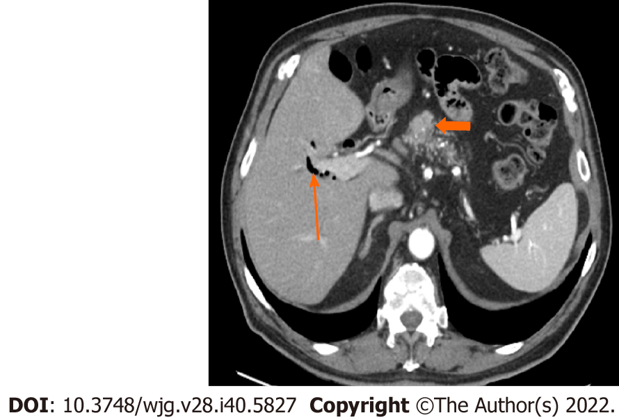
Figure 3 Stage IA (T1, N0, M0).
A 77-year-old male patient with acinar cell carcinoma. Axial post-contrast portal-venous phase computed tomography image shows a solid mass (short arrows) measuring 1.8 cm × 1.6 cm and involving the region of the pancreatic head/body. Incidental findings are calcifications seen throughout the pancreas (likely related to changes in chronic pancreatitis) and mild dilatation of the biliary tree with pneumobilia (long arrow).
- Citation: Calimano-Ramirez LF, Daoud T, Gopireddy DR, Morani AC, Waters R, Gumus K, Klekers AR, Bhosale PR, Virarkar MK. Pancreatic acinar cell carcinoma: A comprehensive review. World J Gastroenterol 2022; 28(40): 5827-5844
- URL: https://www.wjgnet.com/1007-9327/full/v28/i40/5827.htm
- DOI: https://dx.doi.org/10.3748/wjg.v28.i40.5827