
















©The Author(s) 2022.
World J Gastroenterol. Aug 7, 2022; 28(29): 3994-4006
Published online Aug 7, 2022. doi: 10.3748/wjg.v28.i29.3994
Published online Aug 7, 2022. doi: 10.3748/wjg.v28.i29.3994
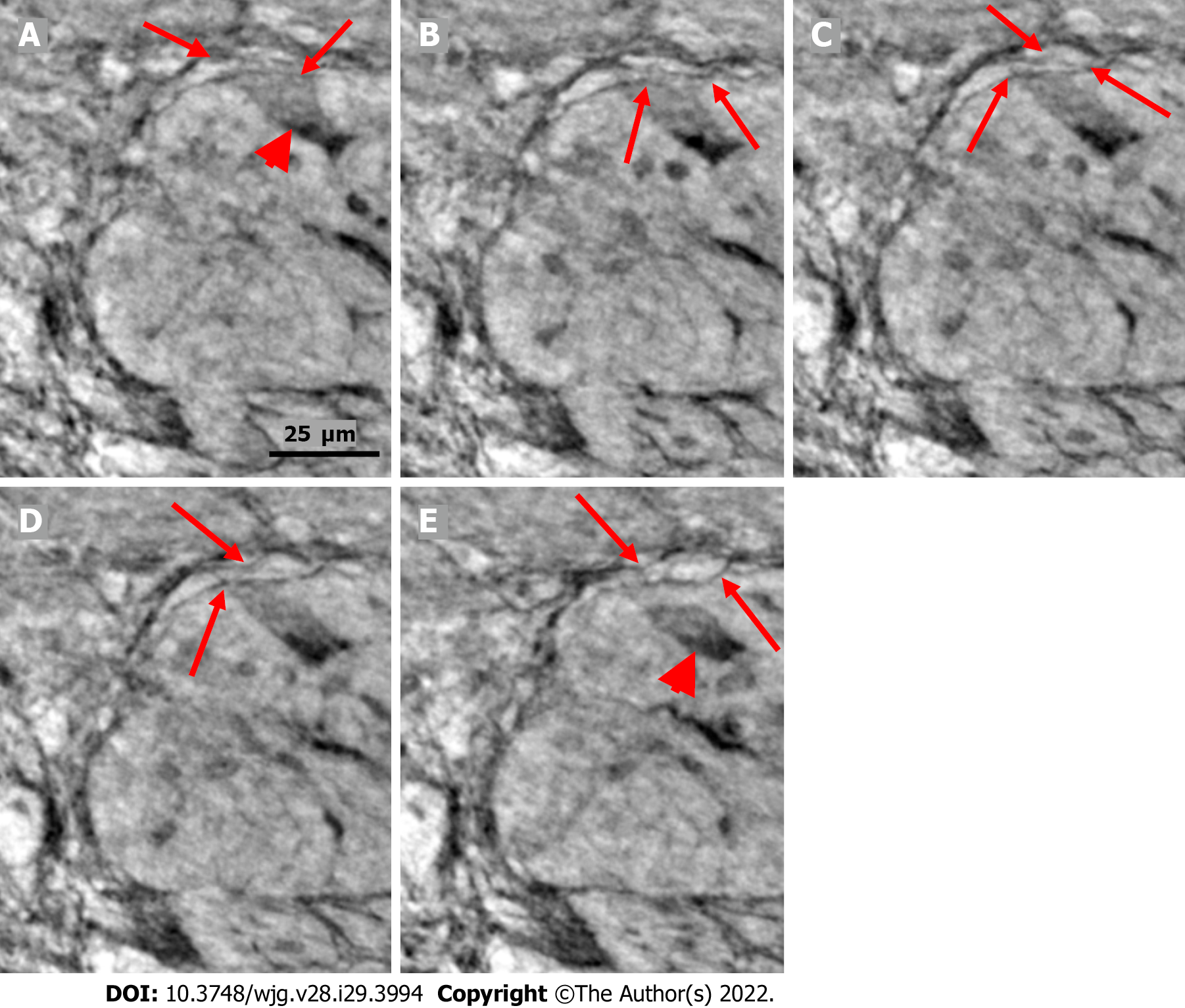
Figure 6 Series of virtual slices covering 6.
4 μm thickness of the ganglion from one patient. A: Neuron with shrunken pyknotic nucleus and dark cytoplasm containing vacuoles at the edge of the ganglion (arrowhead). A part of the cytoplasm of the pre-apoptotic neuron is present between the telopodes (arrows); B: 2.0 μm deeper from Figure A. The lower telopode was discontinuous (arrows); C: 0.7 μm deeper from Figure B. The two telopodes are intact, and almost all of the cytoplasm between them has disappeared; D: 0.7 μm deeper from Figure C. Pre-apoptotic neurons seem to be in contact with the lower telopode; E: 3.0 μm deeper from Figure D. The pre-apoptotic neuron (arrowhead) lies within the ganglion, without contact with the telopodes. Arrows point to two telopodes on the figures. The scale bar (25 μm) applies to all the subfigures.
- Citation: Veress B, Peruzzi N, Eckermann M, Frohn J, Salditt T, Bech M, Ohlsson B. Structure of the myenteric plexus in normal and diseased human ileum analyzed by X-ray virtual histology slices. World J Gastroenterol 2022; 28(29): 3994-4006
- URL: https://www.wjgnet.com/1007-9327/full/v28/i29/3994.htm
- DOI: https://dx.doi.org/10.3748/wjg.v28.i29.3994