
















©The Author(s) 2021.
World J Gastroenterol. Dec 14, 2021; 27(46): 7894-7908
Published online Dec 14, 2021. doi: 10.3748/wjg.v27.i46.7894
Published online Dec 14, 2021. doi: 10.3748/wjg.v27.i46.7894
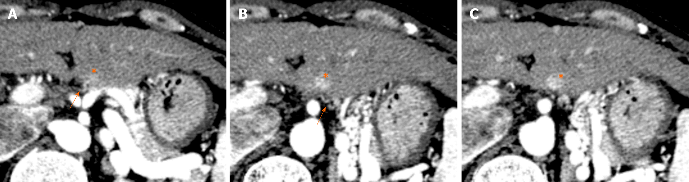
Figure 8 Hypervascular pseudolesion observed in posterior aspect of segment II (40th female).
A-C: On arterial phase images of contrast enhanced computed tomography, tiny focal hyper-attenuation area is observed in posterior aspect of segment II of the liver (*). Tiny vascular branch is directly entering the area from outside of the liver, which is an aberrant left gastric vein directly entering to the liver (arrows).
- Citation: Kobayashi S. Hepatic pseudolesions caused by alterations in intrahepatic hemodynamics. World J Gastroenterol 2021; 27(46): 7894-7908
- URL: https://www.wjgnet.com/1007-9327/full/v27/i46/7894.htm
- DOI: https://dx.doi.org/10.3748/wjg.v27.i46.7894