
















©The Author(s) 2021.
World J Gastroenterol. Jun 14, 2021; 27(22): 3097-3108
Published online Jun 14, 2021. doi: 10.3748/wjg.v27.i22.3097
Published online Jun 14, 2021. doi: 10.3748/wjg.v27.i22.3097
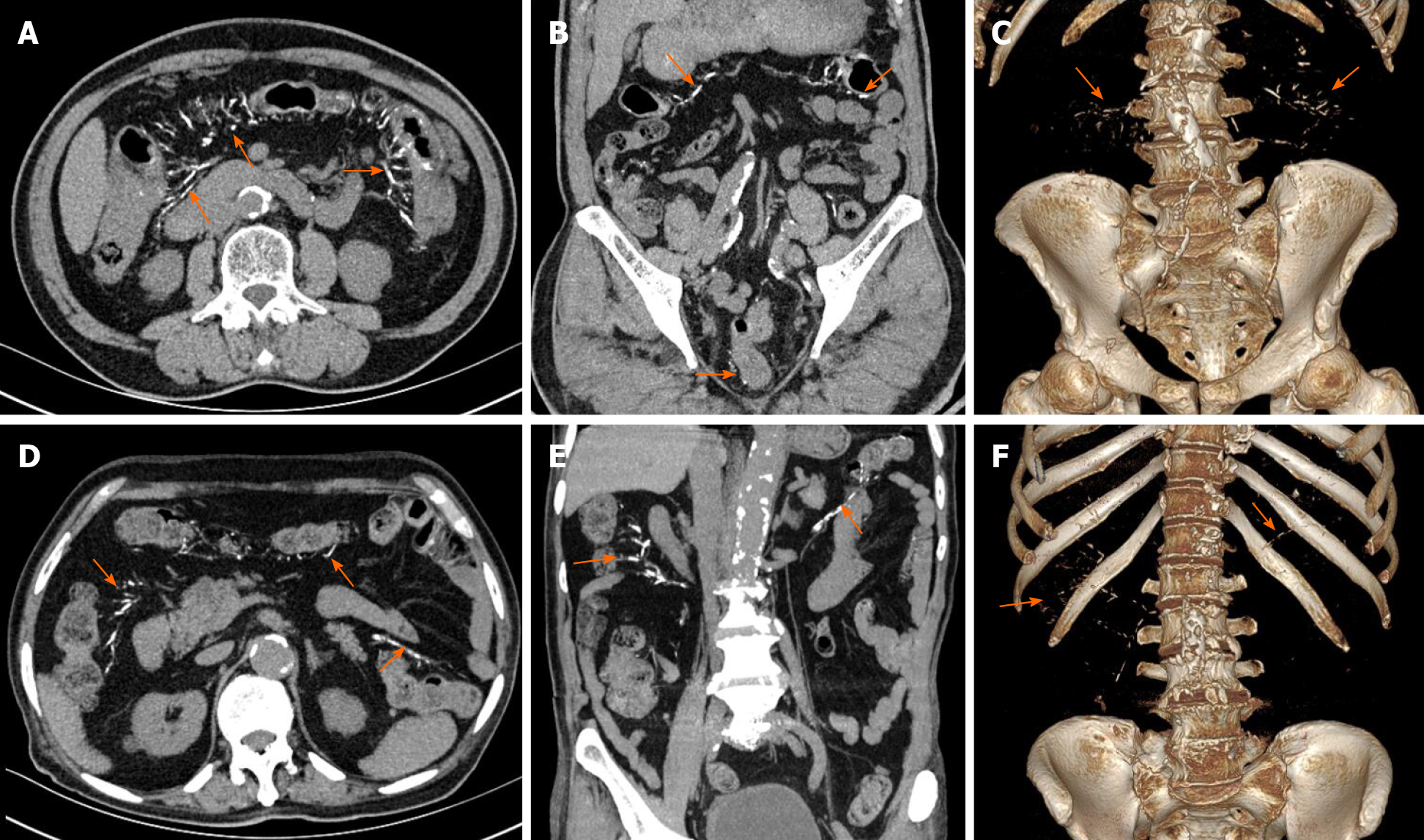
Figure 5 Abdominal computed tomography shows multiple threadlike calcifications within the colon wall and adjacent vein from the ileocecal junction to the descending colon (arrow).
A-C: Case 6; D-F: Case 7. In case 6, calcifications of the mesenteric vein extended to the rectum; mild diffuse thickening of colon wall is seen. Volume rendering image illustrates the distribution of calcifications in the mesenteric veins, the inferior mesenteric vein with multifocal calcifications (C, F).
- Citation: Wen Y, Chen YW, Meng AH, Zhao M, Fang SH, Ma YQ. Idiopathic mesenteric phlebosclerosis associated with long-term oral intake of geniposide. World J Gastroenterol 2021; 27(22): 3097-3108
- URL: https://www.wjgnet.com/1007-9327/full/v27/i22/3097.htm
- DOI: https://dx.doi.org/10.3748/wjg.v27.i22.3097