
















©The Author(s) 2021.
World J Gastroenterol. Apr 28, 2021; 27(16): 1841-1846
Published online Apr 28, 2021. doi: 10.3748/wjg.v27.i16.1841
Published online Apr 28, 2021. doi: 10.3748/wjg.v27.i16.1841
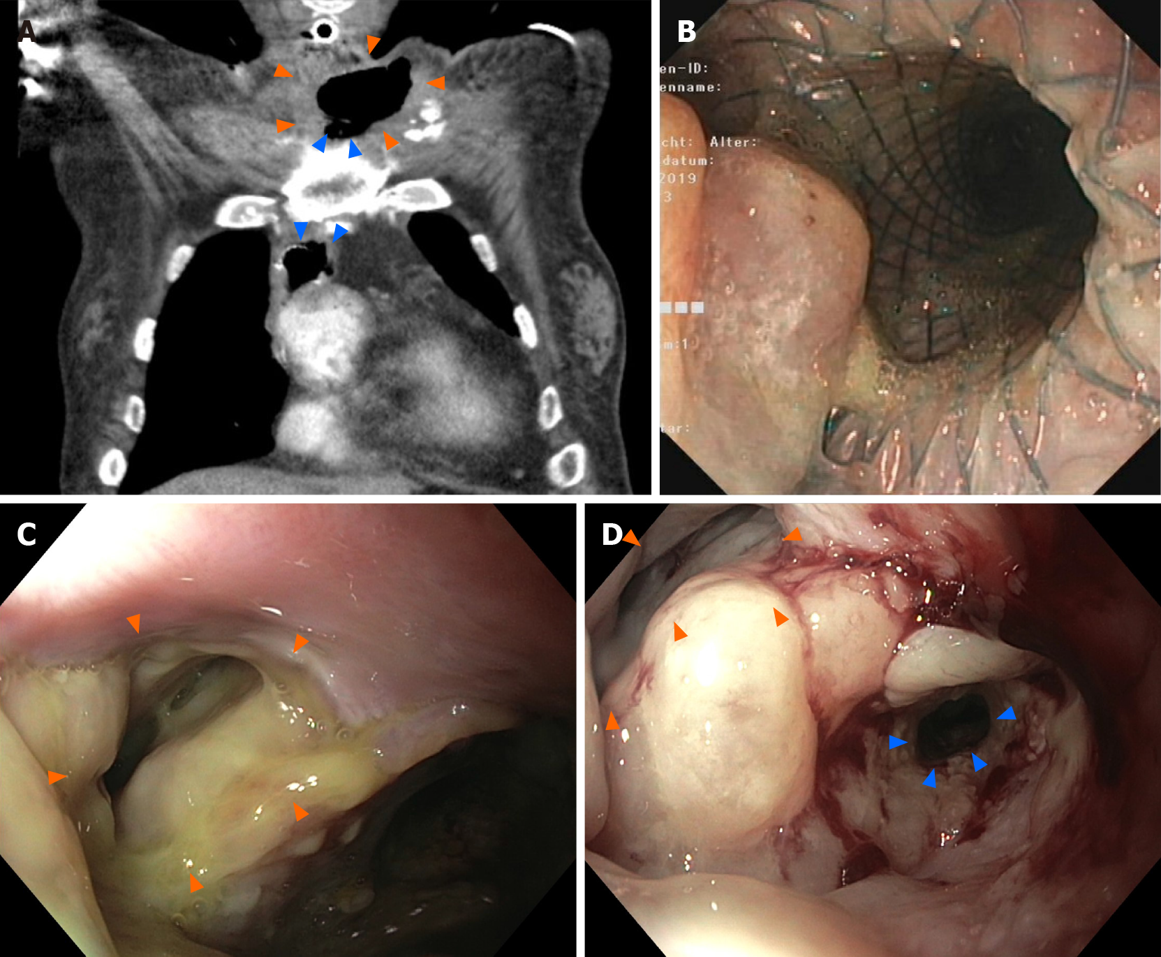
Figure 1 Preoperative findings.
A: Contrast-enhanced computed tomography with oral and intravenous contrast application showing the jugular abscess (indicated by orange arrowheads) and the fully covered self-expandable metallic stent with its proximal end in the fistula (indicated by blue arrowheads); B: Endoscopy showing fully covered self-expandable metallic stent placed in the distal stenotic esophagus with the esophagocutaneous fistula directly orally of the stent. The fistula orifice is partially covered by the stent flare; C: Endoscopy after stent extraction with the large prestenotic fistula orifice (indicated by orange arrowheads); fistula filled with pus; D: Endoscopy after endoscopic vacuum therapy with cleaned fistula (indicated by orange arrowhead). The fistula is entirely epithelized. The distal esophagus segment remains stenotic (indicated by blue arrowheads).
- Citation: Lock JF, Reimer S, Pietryga S, Jakubietz R, Flemming S, Meining A, Germer CT, Seyfried F. Managing esophagocutaneous fistula after secondary gastric pull-up: A case report. World J Gastroenterol 2021; 27(16): 1841-1846
- URL: https://www.wjgnet.com/1007-9327/full/v27/i16/1841.htm
- DOI: https://dx.doi.org/10.3748/wjg.v27.i16.1841