
















©The Author(s) 2020.
World J Gastroenterol. Mar 7, 2020; 26(9): 960-972
Published online Mar 7, 2020. doi: 10.3748/wjg.v26.i9.960
Published online Mar 7, 2020. doi: 10.3748/wjg.v26.i9.960
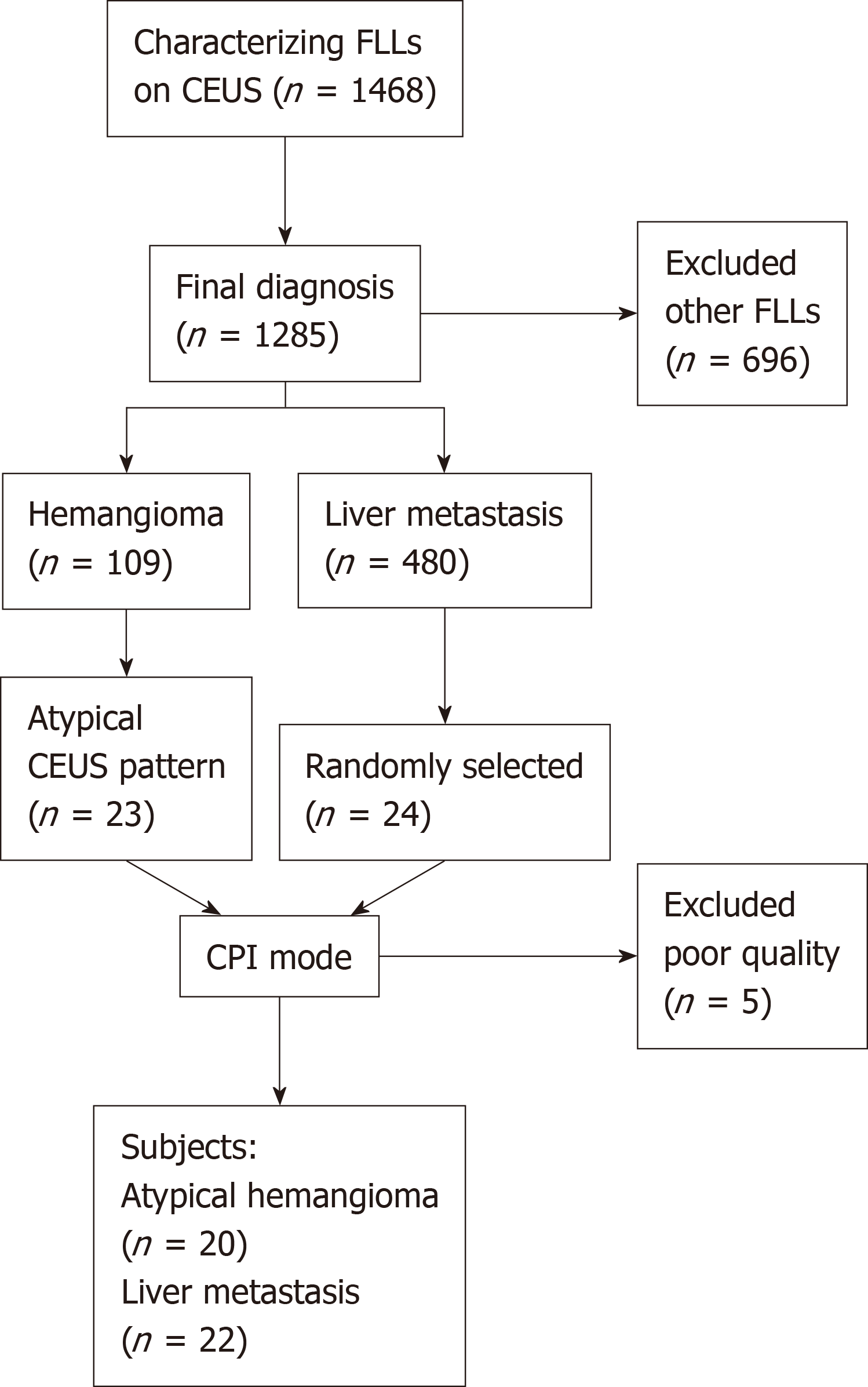
Figure 1 Flow diagram of the study population.
CPI: Color parametric imaging; FLL: Focal liver lesions, CEUS: Contrast enhanced ultrasound.
- Citation: Wu XF, Bai XM, Yang W, Sun Y, Wang H, Wu W, Chen MH, Yan K. Differentiation of atypical hepatic hemangioma from liver metastases: Diagnostic performance of a novel type of color contrast enhanced ultrasound. World J Gastroenterol 2020; 26(9): 960-972
- URL: https://www.wjgnet.com/1007-9327/full/v26/i9/960.htm
- DOI: https://dx.doi.org/10.3748/wjg.v26.i9.960