
















©The Author(s) 2020.
World J Gastroenterol. Oct 7, 2020; 26(37): 5731-5744
Published online Oct 7, 2020. doi: 10.3748/wjg.v26.i37.5731
Published online Oct 7, 2020. doi: 10.3748/wjg.v26.i37.5731
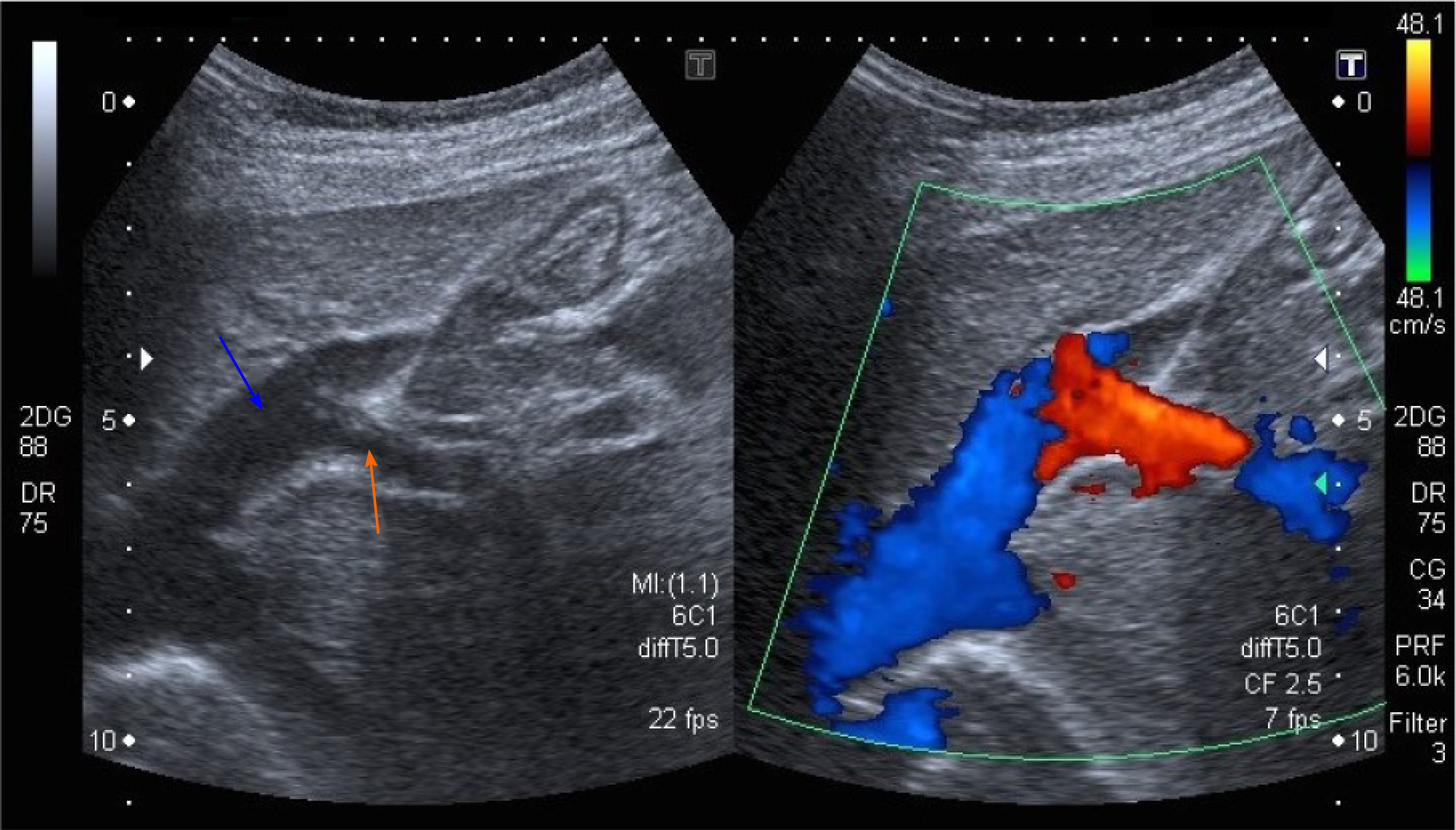
Figure 1 Abdominal ultrasound with Doppler color flow (case 1).
An anomalous vascular connection between the left hepatic vein (blue arrow) and left portal vein (orange arrow) is seen.
- Citation: Peček J, Fister P, Homan M. Abernethy syndrome in Slovenian children: Five case reports and review of literature. World J Gastroenterol 2020; 26(37): 5731-5744
- URL: https://www.wjgnet.com/1007-9327/full/v26/i37/5731.htm
- DOI: https://dx.doi.org/10.3748/wjg.v26.i37.5731