
















©The Author(s) 2019.
World J Gastroenterol. Sep 21, 2019; 25(35): 5334-5343
Published online Sep 21, 2019. doi: 10.3748/wjg.v25.i35.5334
Published online Sep 21, 2019. doi: 10.3748/wjg.v25.i35.5334
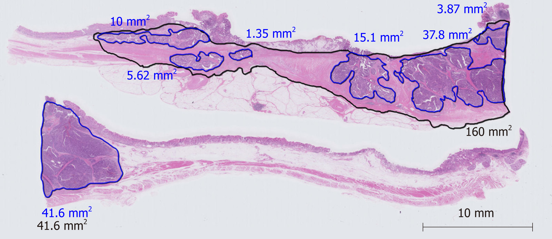
Figure 1 An example of a virtual microscopic slide and pathological diagnosis of the residual tumor area and primary tumorous bed.
The areas enclosed by a red line and a black line are the residual tumor area and the primary tumorous bed, respectively. Their square measures were automatically calculated by the software of the virtual microscopic system.
- Citation: Kawai S, Shimoda T, Nakajima T, Terashima M, Omae K, Machida N, Yasui H. Pathological response measured using virtual microscopic slides for gastric cancer patients who underwent neoadjuvant chemotherapy. World J Gastroenterol 2019; 25(35): 5334-5343
- URL: https://www.wjgnet.com/1007-9327/full/v25/i35/5334.htm
- DOI: https://dx.doi.org/10.3748/wjg.v25.i35.5334