
















©The Author(s) 2018.
World J Gastroenterol. Sep 7, 2018; 24(33): 3806-3812
Published online Sep 7, 2018. doi: 10.3748/wjg.v24.i33.3806
Published online Sep 7, 2018. doi: 10.3748/wjg.v24.i33.3806
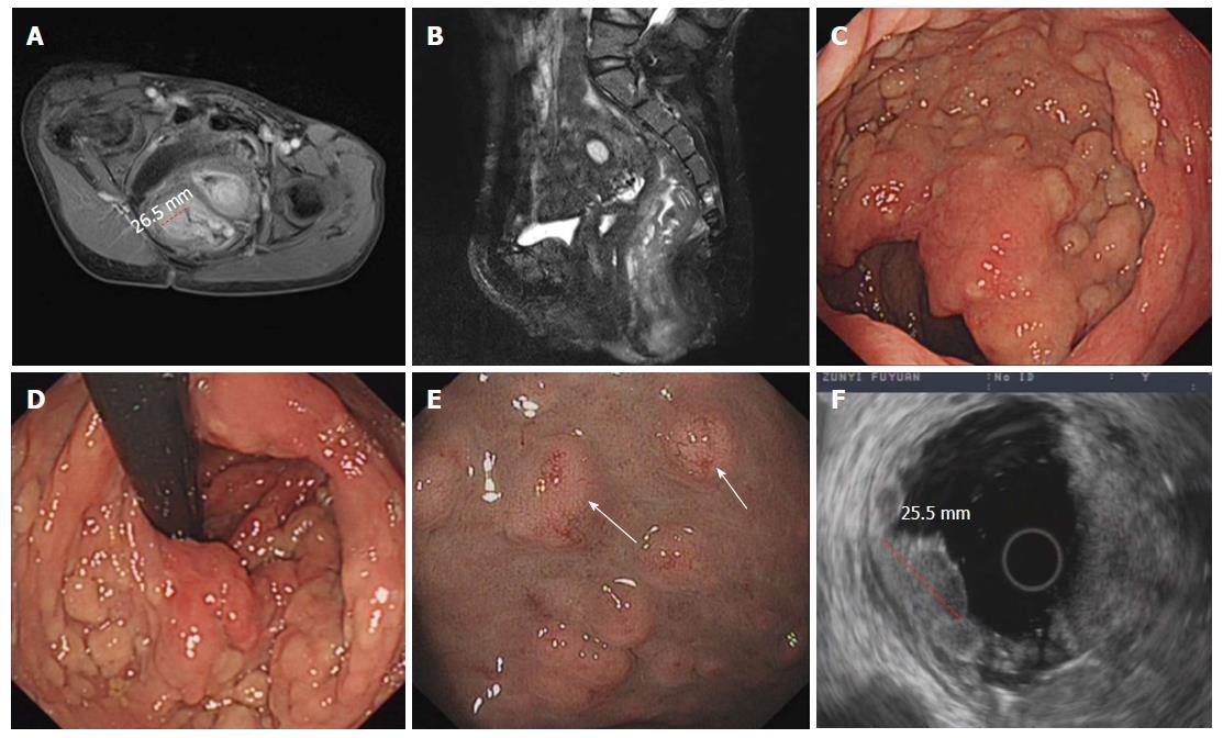
Figure 3 Imaging, endoscopy and endoscopy ultrasonographic findings of multiple rectal neuroendocrine tumors in the patient.
A and B: Magnetic resonance imaging (red dotted line marking the widest point of the tumors, measuring approximately 26.5 mm); C and D: Endoscopic manifestations; E: Blood vessels were apparent on the surface of the nodules under the NBI (neuroendocrine tumors marked by the white arrow); F: Endoscopic ultrasonography of the multiple rectal neuroendocrine tumors (red dotted line marking the widest point of the tumors, measuring approximately 25.5 mm). NBI: Narrow-band imaging.
- Citation: Xie R, Fu KI, Chen SM, Tuo BG, Wu HC. Neurofibromatosis type 1-associated multiple rectal neuroendocrine tumors: A case report and review of the literature. World J Gastroenterol 2018; 24(33): 3806-3812
- URL: https://www.wjgnet.com/1007-9327/full/v24/i33/3806.htm
- DOI: https://dx.doi.org/10.3748/wjg.v24.i33.3806